 Movie
Movie Controller

[English] 日本語
 Yorodumi
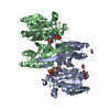
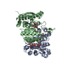
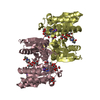
- PDB-6xxy: Crystal structure of Haemophilus influenzae 3-isopropylmalate deh... -
+ Open data

- Basic information
| Entry | Database: PDB / ID: 6xxy | ||||||
|---|---|---|---|---|---|---|---|
| Title | Crystal structure of Haemophilus influenzae 3-isopropylmalate dehydrogenase in complex with O-isobutenyl oxalylhydroxamate. | ||||||
 Components Components | 3-isopropylmalate dehydrogenase | ||||||
 Keywords Keywords | OXIDOREDUCTASE / Haemophilus influenzae / leucine biosynthetis / 3-isopropylmalate dehydrogenase / inhibitor | ||||||
| Function / homology |  Function and homology information Function and homology information3-isopropylmalate dehydrogenase / 3-isopropylmalate dehydrogenase activity / L-leucine biosynthetic process / NAD binding / magnesium ion binding / cytosol Similarity search - Function | ||||||
| Biological species |  Haemophilus influenzae (bacteria) Haemophilus influenzae (bacteria) | ||||||
| Method |  X-RAY DIFFRACTION / SYNCHROTRON / MOLECULAR REPLACEMENT / Resolution: 2.09 Å X-RAY DIFFRACTION / SYNCHROTRON / MOLECULAR REPLACEMENT / Resolution: 2.09 Å | ||||||
 Authors Authors | Miggiano, R. / Rossi, F. / Martignon, S. / Rizzi, M. | ||||||
 Citation Citation | Journal: Biochem.Biophys.Res.Commun. / Year: 2020 Title: Crystal structure of Haemophilus influenzae 3-isopropylmalate dehydrogenase (LeuB) in complex with the inhibitor O-isobutenyl oxalylhydroxamate. Authors: Miggiano, R. / Martignon, S. / Minassi, A. / Rossi, F. / Rizzi, M. | ||||||
| History |
|
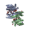
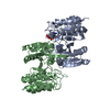
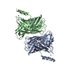
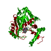
- Structure visualization
| Structure viewer | Molecule: MolmilJmol/JSmol |
|---|
- Downloads & links
-Download
| PDBx/mmCIF format | 6xxy.cif.gz | 196.1 KB | Display | PDBx/mmCIF format |
|---|---|---|---|---|
| PDB format | pdb6xxy.ent.gz | 125.3 KB | Display | PDB format |
| PDBx/mmJSON format | 6xxy.json.gz | Tree view | PDBx/mmJSON format | |
| Others |  Other downloads Other downloads |
-Validation report
| Arichive directory | https://data.pdbj.org/pub/pdb/validation_reports/xx/6xxyftp://data.pdbj.org/pub/pdb/validation_reports/xx/6xxy | HTTPS FTP |
|---|
-Related structure data
| Related structure data | 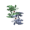 1a05S S: Starting model for refinement |
|---|---|
| Similar structure data |
-Links
 PDBj
PDBj

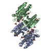
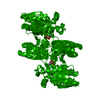
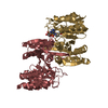
- Assembly
| Deposited unit | 
| ||||||||||||
|---|---|---|---|---|---|---|---|---|---|---|---|---|---|
| 1 |
| ||||||||||||
| Unit cell |
|
-Components
| #1: Protein | Mass: 38847.324 Da / Num. of mol.: 2 / Mutation: Q2E Source method: isolated from a genetically manipulated source Details: Glutammic acid at residue 2 has been introduced as result of subcloning strategies Source: (gene. exp.) Haemophilus influenzae (strain ATCC 51907 / DSM 11121 / KW20 / Rd) (bacteria)Strain: ATCC 51907 / DSM 11121 / KW20 / Rd / Gene: leuB, HI_0987 / Production host: References: UniProt: P43860, 3-isopropylmalate dehydrogenase #2: Chemical |   Mass: 663.425 Da / Num. of mol.: 2 / Source method: obtained synthetically / Formula: C21H27N7O14P2 / Comment: NAD*YM Mass: 663.425 Da / Num. of mol.: 2 / Source method: obtained synthetically / Formula: C21H27N7O14P2 / Comment: NAD*YM#3: Chemical |   Mass: 24.305 Da / Num. of mol.: 2 / Source method: obtained synthetically / Formula: Mg Mass: 24.305 Da / Num. of mol.: 2 / Source method: obtained synthetically / Formula: Mg#4: Chemical | 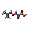  Mass: 159.140 Da / Num. of mol.: 2 / Source method: obtained synthetically / Formula: C6H9NO4 / Feature type: SUBJECT OF INVESTIGATION Mass: 159.140 Da / Num. of mol.: 2 / Source method: obtained synthetically / Formula: C6H9NO4 / Feature type: SUBJECT OF INVESTIGATION#5: Water | ChemComp-HOH / |  Mass: 18.015 Da / Num. of mol.: 353 / Source method: isolated from a natural source / Formula: H2O Mass: 18.015 Da / Num. of mol.: 353 / Source method: isolated from a natural source / Formula: H2OHas ligand of interest | Y | |
|---|
-Experimental details
-Experiment
| Experiment | Method: X-RAY DIFFRACTION / Number of used crystals: 1 |
|---|
- Sample preparation
| Crystal | Density Matthews: 2.35 Å3/Da / Density % sol: 47 % |
|---|---|
| Crystal grow | Temperature: 293.15 K / Method: vapor diffusion, sitting drop Details: 2 M (NH4)2SO4, 0.1 M Na-citrate pH 5.5 and 5% 2-propanol. |
-Data collection
| Diffraction | Mean temperature: 93.15 K / Serial crystal experiment: N |
|---|---|
| Diffraction source | Source: SYNCHROTRON / Site: ESRF  / Beamline: ID29 / Wavelength: 0.976 Å / Beamline: ID29 / Wavelength: 0.976 Å |
| Detector | Type: ADSC QUANTUM 315 / Detector: CCD / Date: Dec 14, 2007 |
| Radiation | Protocol: SINGLE WAVELENGTH / Monochromatic (M) / Laue (L): M / Scattering type: x-ray |
| Radiation wavelength | Wavelength: 0.976 Å / Relative weight: 1 |
| Reflection | Resolution: 2.09→58.49 Å / Num. obs: 41922 / % possible obs: 98.53 % / Redundancy: 1.9 % / Biso Wilson estimate: 24.9 Å2 / Rmerge(I) obs: 0.037 / Rrim(I) all: 0.053 / Net I/σ(I): 12.75 |
| Reflection shell | Resolution: 2.09→2.39 Å / Rmerge(I) obs: 0.2037 / Mean I/σ(I) obs: 4.59 / Num. unique obs: 3733 |
- Processing
| Software |
| ||||||||||||||||||||||||||||
|---|---|---|---|---|---|---|---|---|---|---|---|---|---|---|---|---|---|---|---|---|---|---|---|---|---|---|---|---|---|
| Refinement | Method to determine structure: MOLECULAR REPLACEMENT Starting model: 1A05 Resolution: 2.09→58.49 Å / SU ML: 0.2852 / Cross valid method: FREE R-VALUE / σ(F): 1.35 / Phase error: 24.8791
| ||||||||||||||||||||||||||||
| Solvent computation | Shrinkage radii: 0.9 Å / VDW probe radii: 1.11 Å | ||||||||||||||||||||||||||||
| Displacement parameters | Biso mean: 35.55 Å2 | ||||||||||||||||||||||||||||
| Refinement step | Cycle: LAST / Resolution: 2.09→58.49 Å
| ||||||||||||||||||||||||||||
| Refine LS restraints |
| ||||||||||||||||||||||||||||
| LS refinement shell |
|
