[English] 日本語
Yorodumi
- PDB-6xr0: Crystal Structure of Human Melanotransferrin in complex with SC57... -

+
Open data


ID or keywords:

Loading...

-
Basic information

Entry
Database: PDB / ID: 6xr0
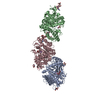
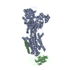
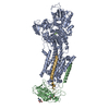
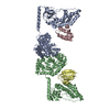
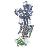
TitleCrystal Structure of Human Melanotransferrin in complex with SC57.32 Fab
Components
  • Melanotransferrin
  • SC57.32 Fab Heavy Chain
  • SC57.32 Fab Light Chain
KeywordsMETAL TRANSPORT / Iron-binding / Ab Complex
Function / homology
Function and homology information


: / protein metabolic process => GO:0019538 / positive regulation of extracellular matrix disassembly / negative regulation of substrate adhesion-dependent cell spreading / : / Post-translational modification: synthesis of GPI-anchored proteins / positive regulation of plasminogen activation / post-translational protein modification / Post-translational protein phosphorylation / recycling endosome ...: / protein metabolic process => GO:0019538 / positive regulation of extracellular matrix disassembly / negative regulation of substrate adhesion-dependent cell spreading / : / Post-translational modification: synthesis of GPI-anchored proteins / positive regulation of plasminogen activation / post-translational protein modification / Post-translational protein phosphorylation / recycling endosome / Regulation of Insulin-like Growth Factor (IGF) transport and uptake by Insulin-like Growth Factor Binding Proteins (IGFBPs) / iron ion transport / early endosome / iron ion binding / endoplasmic reticulum lumen / cell surface / extracellular space / extracellular exosome / extracellular region / plasma membrane
Similarity search - Function
Melanotransferrin / Transferrin-like domain signature 2. / Transferrin family, iron binding site / Transferrin-like domain signature 1. / Transferrin-like domain signature 3. / Transferrin-like domain / Transferrin / Transferrin / Transferrin-like domain profile. / Transferrin
Similarity search - Domain/homology
BICARBONATE ION / : / Melanotransferrin
Similarity search - Component
Biological speciesHomo sapiens (human)
Mus musculus (house mouse)
MethodX-RAY DIFFRACTION / SYNCHROTRON / MOLECULAR REPLACEMENT / Resolution: 3.064 Å
AuthorsHayashi, K. / Longenecker, K.L. / Vivona, S.
CitationJournal: Sci Rep / Year: 2021
Title: Complex of human Melanotransferrin and SC57.32 Fab fragment reveals novel interdomain arrangement with ferric N-lobe and open C-lobe.
Authors: Hayashi, K. / Longenecker, K.L. / Liu, L.Y. / Faust, B. / Prashar, A. / Hampl, J. / Stoll, V. / Vivona, S.
History
DepositionJul 10, 2020Deposition site: RCSB / Processing site: RCSB
Revision 1.0Jan 20, 2021Provider: repository / Type: Initial release
Revision 1.1Oct 18, 2023Group: Data collection / Database references / Refinement description
Category: chem_comp_atom / chem_comp_bond ...chem_comp_atom / chem_comp_bond / database_2 / pdbx_initial_refinement_model
Item: _database_2.pdbx_DOI / _database_2.pdbx_database_accession

-
Structure visualization

Structure viewerMolecule:
MolmilJmol/JSmol

Downloads & links

-
Assembly

Deposited unit
H: SC57.32 Fab Heavy Chain
L: SC57.32 Fab Light Chain
M: Melanotransferrin
hetero molecules


Theoretical massNumber of molelcules
Total (without water)129,04610
Polymers127,8123
Non-polymers1,2347
Water90150
1


  • Idetical with deposited unit
  • defined by author&software
  • Evidence: gel filtration
TypeNameSymmetry operationNumber
identity operation1_555x,y,z1
Buried area7620 Å2
ΔGint-34 kcal/mol
Surface area46980 Å2
MethodPISA
Unit cell
Length a, b, c (Å)57.829, 136.910, 111.135
Angle α, β, γ (deg.)90.000, 91.460, 90.000
Int Tables number4
Space group name H-MP1211

-
Components

-
Protein , 1 types, 1 molecules M

#3: Protein Melanotransferrin / / Melanoma-associated antigen p97


Mass: 80302.453 Da / Num. of mol.: 1
Source method: isolated from a genetically manipulated source
Source: (gene. exp.) Homo sapiens (human) / Gene: MELTF, MAP97, MFI2 / Production host: Cricetulus griseus (Chinese hamster) / References: UniProt: P08582

-
Antibody , 2 types, 2 molecules HL

#1: Antibody SC57.32 Fab Heavy Chain


Mass: 23844.688 Da / Num. of mol.: 1 / Source method: isolated from a natural source / Source: (natural) Mus musculus (house mouse)
#2: Antibody SC57.32 Fab Light Chain


Mass: 23664.986 Da / Num. of mol.: 1 / Source method: isolated from a natural source / Source: (natural) Mus musculus (house mouse)

-
Sugars , 2 types, 3 molecules

#4: Polysaccharide 2-acetamido-2-deoxy-beta-D-glucopyranose-(1-4)-2-acetamido-2-deoxy-beta-D-glucopyranose


Type: oligosaccharide / Mass: 424.401 Da / Num. of mol.: 2
Source method: isolated from a genetically manipulated source
DescriptorTypeProgram
DGlcpNAcb1-4DGlcpNAcb1-Glycam Condensed SequenceGMML 1.0
WURCS=2.0/1,2,1/[a2122h-1b_1-5_2*NCC/3=O]/1-1/a4-b1WURCSPDB2Glycan 1.1.0
[]{[(4+1)][b-D-GlcpNAc]{[(4+1)][b-D-GlcpNAc]{}}}LINUCSPDB-CARE
#5: Sugar ChemComp-NAG / 2-acetamido-2-deoxy-beta-D-glucopyranose / N-acetyl-beta-D-glucosamine / 2-acetamido-2-deoxy-beta-D-glucose / 2-acetamido-2-deoxy-D-glucose / 2-acetamido-2-deoxy-glucose / N-ACETYL-D-GLUCOSAMINE / N-Acetylglucosamine


Type: D-saccharide, beta linking / Mass: 221.208 Da / Num. of mol.: 1
Source method: isolated from a genetically manipulated source
Formula: C8H15NO6
IdentifierTypeProgram
DGlcpNAcbCONDENSED IUPAC CARBOHYDRATE SYMBOLGMML 1.0
N-acetyl-b-D-glucopyranosamineCOMMON NAMEGMML 1.0
b-D-GlcpNAcIUPAC CARBOHYDRATE SYMBOLPDB-CARE 1.0
GlcNAcSNFG CARBOHYDRATE SYMBOLGMML 1.0

-
Non-polymers , 5 types, 54 molecules

#6: Chemical ChemComp-MG / MAGNESIUM ION


Mass: 24.305 Da / Num. of mol.: 1 / Source method: obtained synthetically / Formula: Mg
#7: Chemical ChemComp-NA / SODIUM ION


Mass: 22.990 Da / Num. of mol.: 1 / Source method: obtained synthetically / Formula: Na
#8: Chemical ChemComp-FE / FE (III) ION / Iron


Mass: 55.845 Da / Num. of mol.: 1 / Source method: obtained synthetically / Formula: Fe / Feature type: SUBJECT OF INVESTIGATION
#9: Chemical ChemComp-BCT / BICARBONATE ION / Bicarbonate


Mass: 61.017 Da / Num. of mol.: 1 / Source method: obtained synthetically / Formula: CHO3 / Comment: pH buffer*YM
#10: Water ChemComp-HOH / water / Water


Mass: 18.015 Da / Num. of mol.: 50 / Source method: isolated from a natural source / Formula: H2O

-
Details

Has ligand of interestY

-
Experimental details

-
Experiment

ExperimentMethod: X-RAY DIFFRACTION / Number of used crystals: 1

-
Sample preparation

CrystalDensity Matthews: 3.44 Å3/Da / Density % sol: 64.25 %
Crystal growTemperature: 294 K / Method: vapor diffusion, sitting drop / Details: 100 mM sodium acetate, pH 4.6 and 30% PEG300

-
Data collection

DiffractionMean temperature: 297 K / Serial crystal experiment: N
Diffraction sourceSource: SYNCHROTRON / Site: APS / Beamline: 17-BM / Wavelength: 1 Å
DetectorType: DECTRIS PILATUS 6M / Detector: PIXEL / Date: Jul 20, 2018
RadiationProtocol: SINGLE WAVELENGTH / Monochromatic (M) / Laue (L): M / Scattering type: x-ray
Radiation wavelengthWavelength: 1 Å / Relative weight: 1
ReflectionResolution: 3.06→111.1 Å / Num. obs: 32506 / % possible obs: 99.8 % / Redundancy: 6.7 % / Rpim(I) all: 0.084 / Net I/σ(I): 12.4
Reflection shellResolution: 3.064→3.117 Å / Num. unique obs: 1623 / Rpim(I) all: 0.621

-
Processing

Software
NameVersionClassification
Aimlessdata scaling
BUSTER2.11.7 (19-MAR-2020)refinement
PDB_EXTRACT3.25data extraction
XDSdata reduction
PHASERphasing
RefinementMethod to determine structure: MOLECULAR REPLACEMENT
Starting model: 1B1X
Resolution: 3.064→111.1 Å / Cor.coef. Fo:Fc: 0.894 / Cor.coef. Fo:Fc free: 0.883 / Cross valid method: THROUGHOUT / σ(F): 0 / SU Rfree Blow DPI: 0.342
RfactorNum. reflection% reflectionSelection details
Rfree0.2249 1728 5.32 %RANDOM
Rwork0.2021 ---
obs0.2033 32506 99.8 %-
Displacement parametersBiso max: 130.9 Å2 / Biso mean: 71.47 Å2 / Biso min: 28.17 Å2
Baniso -1Baniso -2Baniso -3
1-17.663 Å20 Å2-12.3373 Å2
2---18.7958 Å20 Å2
3---1.1327 Å2
Refine analyzeLuzzati coordinate error obs: 0.39 Å
Refinement stepCycle: final / Resolution: 3.064→111.1 Å
ProteinNucleic acidLigandSolventTotal
Num. atoms8545 0 77 50 8672
Biso mean--67.2 48.88 -
Num. residues----1112
Refine LS restraints
Refine-IDTypeNumberRestraint functionWeightDev ideal
X-RAY DIFFRACTIONt_dihedral_angle_d3005SINUSOIDAL2
X-RAY DIFFRACTIONt_trig_c_planes
X-RAY DIFFRACTIONt_gen_planes1523HARMONIC5
X-RAY DIFFRACTIONt_it8873HARMONIC10
X-RAY DIFFRACTIONt_nbd
X-RAY DIFFRACTIONt_improper_torsion
X-RAY DIFFRACTIONt_pseud_angle
X-RAY DIFFRACTIONt_chiral_improper_torsion1157SEMIHARMONIC5
X-RAY DIFFRACTIONt_sum_occupancies
X-RAY DIFFRACTIONt_utility_distance
X-RAY DIFFRACTIONt_utility_angle
X-RAY DIFFRACTIONt_utility_torsion
X-RAY DIFFRACTIONt_ideal_dist_contact6947SEMIHARMONIC4
X-RAY DIFFRACTIONt_bond_d8873HARMONIC20.008
X-RAY DIFFRACTIONt_angle_deg12074HARMONIC20.93
X-RAY DIFFRACTIONt_omega_torsion3.47
X-RAY DIFFRACTIONt_other_torsion20.63
LS refinement shellResolution: 3.064→3.08 Å / Rfactor Rfree error: 0
RfactorNum. reflection% reflection
Rfree0.2982 23 3.53 %
Rwork0.296 628 -
obs--99.08 %

+
About Yorodumi

-
News

-
Feb 9, 2022. New format data for meta-information of EMDB entries

New format data for meta-information of EMDB entries

  • Version 3 of the EMDB header file is now the official format.
  • The previous official version 1.9 will be removed from the archive.

Related info.:EMDB header

External links:wwPDB to switch to version 3 of the EMDB data model

-
Aug 12, 2020. Covid-19 info

Covid-19 info

URL: https://pdbj.org/emnavi/covid19.php

New page: Covid-19 featured information page in EM Navigator.

Related info.:Covid-19 info / Mar 5, 2020. Novel coronavirus structure data

+
Mar 5, 2020. Novel coronavirus structure data

Novel coronavirus structure data

Related info.:Yorodumi Speices / Aug 12, 2020. Covid-19 info

External links:COVID-19 featured content - PDBj / Molecule of the Month (242):Coronavirus Proteases

+
Jan 31, 2019. EMDB accession codes are about to change! (news from PDBe EMDB page)

EMDB accession codes are about to change! (news from PDBe EMDB page)

  • The allocation of 4 digits for EMDB accession codes will soon come to an end. Whilst these codes will remain in use, new EMDB accession codes will include an additional digit and will expand incrementally as the available range of codes is exhausted. The current 4-digit format prefixed with “EMD-” (i.e. EMD-XXXX) will advance to a 5-digit format (i.e. EMD-XXXXX), and so on. It is currently estimated that the 4-digit codes will be depleted around Spring 2019, at which point the 5-digit format will come into force.
  • The EM Navigator/Yorodumi systems omit the EMD- prefix.

Related info.:Q: What is EMD? / ID/Accession-code notation in Yorodumi/EM Navigator

External links:EMDB Accession Codes are Changing Soon! / Contact to PDBj

+
Jul 12, 2017. Major update of PDB

Major update of PDB

  • wwPDB released updated PDB data conforming to the new PDBx/mmCIF dictionary.
  • This is a major update changing the version number from 4 to 5, and with Remediation, in which all the entries are updated.
  • In this update, many items about electron microscopy experimental information are reorganized (e.g. em_software).
  • Now, EM Navigator and Yorodumi are based on the updated data.

External links:wwPDB Remediation / Enriched Model Files Conforming to OneDep Data Standards Now Available in the PDB FTP Archive

-
Yorodumi

Thousand views of thousand structures

  • Yorodumi is a browser for structure data from EMDB, PDB, SASBDB, etc.
  • This page is also the successor to EM Navigator detail page, and also detail information page/front-end page for Omokage search.
  • The word "yorodu" (or yorozu) is an old Japanese word meaning "ten thousand". "mi" (miru) is to see.

Related info.:EMDB / PDB / SASBDB / Comparison of 3 databanks / Yorodumi Search / Aug 31, 2016. New EM Navigator & Yorodumi / Yorodumi Papers / Jmol/JSmol / Function and homology information / Changes in new EM Navigator and Yorodumi

Read more