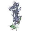
Journal: Proc Natl Acad Sci U S A / Year: 2012 Title: Cryo-EM structure of gastric H+,K+-ATPase with a single occupied cation-binding site. Authors: Kazuhiro Abe / Kazutoshi Tani / Thomas Friedrich / Yoshinori Fujiyoshi / Abstract: Gastric H(+),K(+)-ATPase is responsible for gastric acid secretion. ATP-driven H(+) uptake into the stomach is efficiently accomplished by the exchange of an equal amount of K(+), resulting in a ...Gastric H(+),K(+)-ATPase is responsible for gastric acid secretion. ATP-driven H(+) uptake into the stomach is efficiently accomplished by the exchange of an equal amount of K(+), resulting in a luminal pH close to 1. Because of the limited free energy available for ATP hydrolysis, the stoichiometry of transported cations is thought to vary from 2H(+)/2K(+) to 1H(+)/1K(+) per hydrolysis of one ATP molecule as the luminal pH decreases, although direct evidence for this hypothesis has remained elusive. Here, we show, using the phosphate analog aluminum fluoride (AlF) and a K(+) congener (Rb(+)), the 8-Å resolution structure of H(+),K(+)-ATPase in the transition state of dephosphorylation, (Rb(+))E2~AlF, which is distinct from the preceding Rb(+)-free E2P state. A strong density located in the transmembrane cation-binding site of (Rb(+))E2~AlF highly likely represents a single bound Rb(+) ion, which is clearly different from the Rb(+)-free E2AlF or K(+)-bound (K(+))E2~AlF structures. Measurement of radioactive (86)Rb(+) binding suggests that the binding stoichiometry varies depending on the pH, and approximately half of the amount of Rb(+) is bound under acidic crystallization conditions compared with at a neutral pH. These data represent structural and biochemical evidence for the 1H(+)/1K(+)/1ATP transport mode of H(+),K(+)-ATPase, which is a prerequisite for generation of the 10(6)-fold proton gradient in terms of thermodynamics. Together with the released E2P-stabilizing interaction between the β subunit's N terminus and the P domain observed in the (Rb(+))E2~AlF structure, we propose a refined vectorial transport model of H(+),K(+)-ATPase, which must prevail against the highly acidic state of the gastric lumen.
History
Deposition
Oct 13, 2012
Deposition site: PDBE / Processing site: PDBE
Revision 1.0
Nov 7, 2012
Provider: repository / Type: Initial release
Revision 1.1
Jan 16, 2013
Group: Database references / Derived calculations / Other
Embedding applied: NO / Shadowing applied: NO / Staining applied: NO / Vitrification applied: YES
Vitrification
Instrument: LEICA KF80 / Cryogen name: NITROGEN
Crystal grow
pH: 4.8 / Details: pH 4.8
-
Data collection
Microscopy
Model: JEOL KYOTO-3000SFF / Date: Mar 23, 2010
Electron gun
Electron source: FIELD EMISSION GUN / Accelerating voltage: 300 kV / Illumination mode: FLOOD BEAM
Electron lens
Mode: BRIGHT FIELD / Nominal magnification: 40000 X / Nominal defocus max: 3480 nm / Nominal defocus min: 830 nm
Image recording
Film or detector model: KODAK SO-163 FILM
Diffraction
Mean temperature: 4 K
Detector
Date: Mar 23, 2010
Radiation
Scattering type: electron
Radiation wavelength
Relative weight: 1
Reflection
Resolution: 8→129 Å / Num. obs: 39197 / % possible obs: 73.2 %
-
Processing
Software
Name
Version
Classification
MRC
modelbuilding
SITUS
refinement
MRC
SUITE
datascaling
MRC
phasing
3D reconstruction
Resolution: 8 Å / Symmetry type: 2D CRYSTAL
Refinement
Resolution: 8→129 Å / Num. reflection obs: 4166 / σ(F): 0 Details: ALL REGIONS WERE MODELED STEREOCHEMICALLY. SUBMISSION BASED ON EXPERIMENTAL DATA FROM EMDB EMD-2219. (DEPOSITION ID: 11197).
Refinement step
Cycle: LAST / Resolution: 8→129 Å
Protein
Nucleic acid
Ligand
Solvent
Total
Num. atoms
9694
0
0
0
9694
Refine LS restraints
Refine-ID
Type
Dev ideal
ELECTRONCRYSTALLOGRAPHY
o_bond_d
0.018
ELECTRONCRYSTALLOGRAPHY
o_bond_d_na
ELECTRONCRYSTALLOGRAPHY
o_bond_d_prot
ELECTRONCRYSTALLOGRAPHY
o_angle_d
ELECTRONCRYSTALLOGRAPHY
o_angle_d_na
ELECTRONCRYSTALLOGRAPHY
o_angle_d_prot
ELECTRONCRYSTALLOGRAPHY
o_angle_deg
2.1
ELECTRONCRYSTALLOGRAPHY
o_angle_deg_na
ELECTRONCRYSTALLOGRAPHY
o_angle_deg_prot
ELECTRONCRYSTALLOGRAPHY
o_dihedral_angle_d
ELECTRONCRYSTALLOGRAPHY
o_dihedral_angle_d_na
ELECTRONCRYSTALLOGRAPHY
o_dihedral_angle_d_prot
ELECTRONCRYSTALLOGRAPHY
o_improper_angle_d
ELECTRONCRYSTALLOGRAPHY
o_improper_angle_d_na
ELECTRONCRYSTALLOGRAPHY
o_improper_angle_d_prot
ELECTRONCRYSTALLOGRAPHY
o_mcbond_it
ELECTRONCRYSTALLOGRAPHY
o_mcangle_it
ELECTRONCRYSTALLOGRAPHY
o_scbond_it
ELECTRONCRYSTALLOGRAPHY
o_scangle_it
+
About Yorodumi
-
News
-
Feb 9, 2022. New format data for meta-information of EMDB entries
New format data for meta-information of EMDB entries
Version 3 of the EMDB header file is now the official format.
The previous official version 1.9 will be removed from the archive.
In the structure databanks used in Yorodumi, some data are registered as the other names, "COVID-19 virus" and "2019-nCoV". Here are the details of the virus and the list of structure data.
Jan 31, 2019. EMDB accession codes are about to change! (news from PDBe EMDB page)
EMDB accession codes are about to change! (news from PDBe EMDB page)
The allocation of 4 digits for EMDB accession codes will soon come to an end. Whilst these codes will remain in use, new EMDB accession codes will include an additional digit and will expand incrementally as the available range of codes is exhausted. The current 4-digit format prefixed with “EMD-” (i.e. EMD-XXXX) will advance to a 5-digit format (i.e. EMD-XXXXX), and so on. It is currently estimated that the 4-digit codes will be depleted around Spring 2019, at which point the 5-digit format will come into force.
The EM Navigator/Yorodumi systems omit the EMD- prefix.
Related info.:Q: What is EMD? / ID/Accession-code notation in Yorodumi/EM Navigator
Yorodumi is a browser for structure data from EMDB, PDB, SASBDB, etc.
This page is also the successor to EM Navigator detail page, and also detail information page/front-end page for Omokage search.
The word "yorodu" (or yorozu) is an old Japanese word meaning "ten thousand". "mi" (miru) is to see.
Related info.:EMDB / PDB / SASBDB / Comparison of 3 databanks / Yorodumi Search / Aug 31, 2016. New EM Navigator & Yorodumi / Yorodumi Papers / Jmol/JSmol / Function and homology information / Changes in new EM Navigator and Yorodumi
 Movie
Movie Controller


 Open data
 Basic information
 Components
 Keywords
 Function and homology information
 Authors
 Citation
 Structure visualization
 Downloads & links
 Other downloads


 PDBj
PDBj

 Assembly
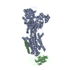
 Sample preparation
 FIELD EMISSION GUN / Accelerating voltage: 300 kV / Illumination mode: FLOOD BEAM
 Processing