 Movie
Movie Controller


+ Open data

- Basic information
| Entry | Database: PDB / ID: 6xnt | ||||||
|---|---|---|---|---|---|---|---|
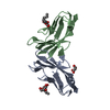
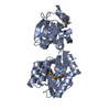
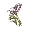
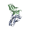
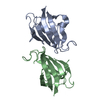
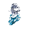
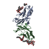
| Title | Crystal structure of I91A mutant of human CEACAM1 | ||||||
 Components Components | Carcinoembryonic antigen-related cell adhesion molecule 1 | ||||||
 Keywords Keywords | IMMUNE SYSTEM / CEACAM1 | ||||||
| Function / homology |  Function and homology information Function and homology informationregulation of endothelial cell differentiation / insulin receptor internalization / negative regulation of cytotoxic T cell degranulation / granulocyte colony-stimulating factor signaling pathway / regulation of homophilic cell adhesion / Regulation of MITF-M dependent genes involved in invasion / negative regulation of hepatocyte proliferation / regulation of epidermal growth factor receptor signaling pathway / regulation of blood vessel remodeling / regulation of sprouting angiogenesis ...regulation of endothelial cell differentiation / insulin receptor internalization / negative regulation of cytotoxic T cell degranulation / granulocyte colony-stimulating factor signaling pathway / regulation of homophilic cell adhesion / Regulation of MITF-M dependent genes involved in invasion / negative regulation of hepatocyte proliferation / regulation of epidermal growth factor receptor signaling pathway / regulation of blood vessel remodeling / regulation of sprouting angiogenesis / filamin binding / negative regulation of natural killer cell mediated cytotoxicity directed against tumor cell target / negative regulation of lipid biosynthetic process / negative regulation of T cell mediated cytotoxicity / regulation of endothelial cell migration / insulin catabolic process / negative regulation of granulocyte differentiation / common myeloid progenitor cell proliferation / negative regulation of interleukin-1 production / regulation of phosphatidylinositol 3-kinase/protein kinase B signal transduction / positive regulation of vasculogenesis / Fibronectin matrix formation / : / negative regulation of fatty acid biosynthetic process / negative regulation of platelet aggregation / bile acid transmembrane transporter activity / regulation of immune system process / wound healing, spreading of cells / negative regulation of vascular permeability / bile acid and bile salt transport / transport vesicle membrane / blood vessel development / microvillus membrane / homophilic cell-cell adhesion / tertiary granule membrane / lateral plasma membrane / negative regulation of T cell receptor signaling pathway / regulation of ERK1 and ERK2 cascade / negative regulation of protein kinase activity / specific granule membrane / regulation of cell migration / basal plasma membrane / protein tyrosine kinase binding / Cell surface interactions at the vascular wall / integrin-mediated signaling pathway / adherens junction / regulation of cell growth / kinase binding / cellular response to insulin stimulus / cell-cell junction / cell junction / cell migration / actin binding / angiogenesis / protein phosphatase binding / calmodulin binding / cell adhesion / protein dimerization activity / apical plasma membrane / Neutrophil degranulation / cell surface / signal transduction / protein homodimerization activity / extracellular exosome / membrane / identical protein binding / plasma membrane Similarity search - Function | ||||||
| Biological species |  Homo sapiens (human) Homo sapiens (human) | ||||||
| Method |  X-RAY DIFFRACTION / SYNCHROTRON / MOLECULAR REPLACEMENT / Resolution: 3.1 Å X-RAY DIFFRACTION / SYNCHROTRON / MOLECULAR REPLACEMENT / Resolution: 3.1 Å | ||||||
 Authors Authors | Gandhi, A.K. / Kim, W.M. / Sun, Z.-Y. / Huang, Y.H. / Bonsor, D. / Petsko, G.A. / Kuchroo, V. / Blumberg, R.S. | ||||||
| Funding support |  United States, 1items United States, 1items
| ||||||
 Citation Citation | Journal: Commun Biol / Year: 2021 Title: Structural basis of the dynamic human CEACAM1 monomer-dimer equilibrium. Authors: Gandhi, A.K. / Sun, Z.J. / Kim, W.M. / Huang, Y.H. / Kondo, Y. / Bonsor, D.A. / Sundberg, E.J. / Wagner, G. / Kuchroo, V.K. / Petsko, G.A. / Blumberg, R.S. | ||||||
| History |
|
- Structure visualization
| Structure viewer | Molecule: MolmilJmol/JSmol |
|---|
- Downloads & links
-Download
| PDBx/mmCIF format | 6xnt.cif.gz | 56.8 KB | Display | PDBx/mmCIF format |
|---|---|---|---|---|
| PDB format | pdb6xnt.ent.gz | 39.9 KB | Display | PDB format |
| PDBx/mmJSON format | 6xnt.json.gz | Tree view | PDBx/mmJSON format | |
| Others |  Other downloads Other downloads |
-Validation report
| Arichive directory | https://data.pdbj.org/pub/pdb/validation_reports/xn/6xntftp://data.pdbj.org/pub/pdb/validation_reports/xn/6xnt | HTTPS FTP |
|---|
-Related structure data
| Related structure data |  6xnoC  6xnwC  6xo1C  4qxwS C: citing same article ( S: Starting model for refinement |
|---|---|
| Similar structure data |


-Links
 PDBj
PDBj


- Assembly
| Deposited unit | 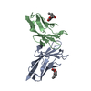
| ||||||||
|---|---|---|---|---|---|---|---|---|---|
| 1 |
| ||||||||
| Unit cell |
|
-Components
| #1: Protein | Mass: 11857.090 Da / Num. of mol.: 2 / Mutation: I91A Source method: isolated from a genetically manipulated source Source: (gene. exp.) Homo sapiens (human) / Gene: CEACAM1, BGP, BGP1 / Production host:  #2: Sugar |   Type: D-saccharide / Mass: 292.369 Da / Num. of mol.: 2 Type: D-saccharide / Mass: 292.369 Da / Num. of mol.: 2Source method: isolated from a genetically manipulated source Formula: C14H28O6 / Comment: detergent*YM #3: Water | ChemComp-HOH / |  Mass: 18.015 Da / Num. of mol.: 4 / Source method: isolated from a natural source / Formula: H2O Mass: 18.015 Da / Num. of mol.: 4 / Source method: isolated from a natural source / Formula: H2OHas ligand of interest | N | |
|---|
-Experimental details
-Experiment
| Experiment | Method: X-RAY DIFFRACTION / Number of used crystals: 1 |
|---|
- Sample preparation
| Crystal | Density Matthews: 3.35 Å3/Da / Density % sol: 63.32 % |
|---|---|
| Crystal grow | Temperature: 298 K / Method: vapor diffusion, sitting drop / pH: 8 Details: 54% Tascimate with 0.5 % n-Octyl-D-glucoside pH 8.0 |
-Data collection
| Diffraction | Mean temperature: 100 K / Serial crystal experiment: N | ||||||||||||||||||||||||||||||
|---|---|---|---|---|---|---|---|---|---|---|---|---|---|---|---|---|---|---|---|---|---|---|---|---|---|---|---|---|---|---|---|
| Diffraction source | Source: SYNCHROTRON / Site: APS / Beamline: 21-ID-G / Wavelength: 0.97856 Å | ||||||||||||||||||||||||||||||
| Detector | Type: MARMOSAIC 300 mm CCD / Detector: CCD / Date: Feb 7, 2017 | ||||||||||||||||||||||||||||||
| Radiation | Protocol: SINGLE WAVELENGTH / Monochromatic (M) / Laue (L): M / Scattering type: x-ray | ||||||||||||||||||||||||||||||
| Radiation wavelength | Wavelength: 0.97856 Å / Relative weight: 1 | ||||||||||||||||||||||||||||||
| Reflection | Resolution: 3.1→72.2 Å / Num. obs: 6265 / % possible obs: 100 % / Redundancy: 11.7 % / CC1/2: 0.963 / Rmerge(I) obs: 0.251 / Rpim(I) all: 0.077 / Rrim(I) all: 0.263 / Net I/σ(I): 9.4 / Num. measured all: 73320 / Scaling rejects: 95 | ||||||||||||||||||||||||||||||
| Reflection shell | Diffraction-ID: 1
|
- Processing
| Software |
| ||||||||||||||||||||||||||||||||||||||||||||||||||||||||||||
|---|---|---|---|---|---|---|---|---|---|---|---|---|---|---|---|---|---|---|---|---|---|---|---|---|---|---|---|---|---|---|---|---|---|---|---|---|---|---|---|---|---|---|---|---|---|---|---|---|---|---|---|---|---|---|---|---|---|---|---|---|---|
| Refinement | Method to determine structure: MOLECULAR REPLACEMENT Starting model: 4QXW Resolution: 3.1→72.2 Å / Cor.coef. Fo:Fc: 0.833 / Cor.coef. Fo:Fc free: 0.848 / SU B: 16.549 / SU ML: 0.293 / Cross valid method: THROUGHOUT / σ(F): 0 / ESU R Free: 0.439 / Stereochemistry target values: MAXIMUM LIKELIHOOD Details: HYDROGENS HAVE BEEN ADDED IN THE RIDING POSITIONS U VALUES : REFINED INDIVIDUALLY
| ||||||||||||||||||||||||||||||||||||||||||||||||||||||||||||
| Solvent computation | Ion probe radii: 0.8 Å / Shrinkage radii: 0.8 Å / VDW probe radii: 1.2 Å / Solvent model: MASK | ||||||||||||||||||||||||||||||||||||||||||||||||||||||||||||
| Displacement parameters | Biso max: 84.99 Å2 / Biso mean: 42.332 Å2 / Biso min: 25.37 Å2
| ||||||||||||||||||||||||||||||||||||||||||||||||||||||||||||
| Refinement step | Cycle: final / Resolution: 3.1→72.2 Å
| ||||||||||||||||||||||||||||||||||||||||||||||||||||||||||||
| Refine LS restraints |
| ||||||||||||||||||||||||||||||||||||||||||||||||||||||||||||
| LS refinement shell | Resolution: 3.1→3.18 Å / Rfactor Rfree error: 0 /
|
