 Movie
Movie Controller

[English] 日本語
 Yorodumi
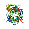
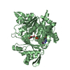
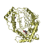
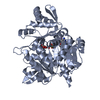
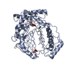
- PDB-6xio: ADP-dependent kinase complex with fructose-6-phosphate and ADPbetaS -
+ Open data

- Basic information
| Entry | Database: PDB / ID: 6xio | ||||||||||||
|---|---|---|---|---|---|---|---|---|---|---|---|---|---|
| Title | ADP-dependent kinase complex with fructose-6-phosphate and ADPbetaS | ||||||||||||
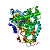
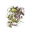
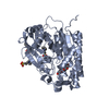
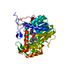
 Components Components | ADP-dependent phosphofructokinase | ||||||||||||
 Keywords Keywords | TRANSFERASE / ancestral protein reconstruction / ADP-dependent sugar kinases family / glucokinase / phosphofructokinase / enzyme evolution | ||||||||||||
| Function / homology | Chem-AT4 / 6-O-phosphono-beta-D-fructofuranose Function and homology information Function and homology information | ||||||||||||
| Biological species |  Methanosarcinales archaeon (archaea) Methanosarcinales archaeon (archaea) | ||||||||||||
| Method |  X-RAY DIFFRACTION / SYNCHROTRON / MOLECULAR REPLACEMENT / Resolution: 3.12 Å X-RAY DIFFRACTION / SYNCHROTRON / MOLECULAR REPLACEMENT / Resolution: 3.12 Å | ||||||||||||
 Authors Authors | Munoz, S. / Gonzalez-Ordenes, F. / Fuentes, N. / Maturana, P. / Herrera-Morande, A. / Villalobos, P. / Castro-Fernandez, V. | ||||||||||||
| Funding support |  Chile, 3items Chile, 3items
| ||||||||||||
 Citation Citation | Journal: J.Biol.Chem. / Year: 2020 Title: Structure of an ancestral ADP-dependent kinase with fructose-6P reveals key residues for binding, catalysis, and ligand-induced conformational changes. Authors: Munoz, S.M. / Castro-Fernandez, V. / Guixe, V. | ||||||||||||
| History |
|
- Structure visualization
| Structure viewer | Molecule: MolmilJmol/JSmol |
|---|
- Downloads & links
-Download
| PDBx/mmCIF format | 6xio.cif.gz | 110.1 KB | Display | PDBx/mmCIF format |
|---|---|---|---|---|
| PDB format | pdb6xio.ent.gz | 78.2 KB | Display | PDB format |
| PDBx/mmJSON format | 6xio.json.gz | Tree view | PDBx/mmJSON format | |
| Others |  Other downloads Other downloads |
-Validation report
| Arichive directory | https://data.pdbj.org/pub/pdb/validation_reports/xi/6xioftp://data.pdbj.org/pub/pdb/validation_reports/xi/6xio | HTTPS FTP |
|---|
-Related structure data
| Related structure data | 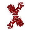 6c8zS S: Starting model for refinement |
|---|---|
| Similar structure data |

-Links
 PDBj
PDBj
- Assembly
| Deposited unit | 
| ||||||||||||
|---|---|---|---|---|---|---|---|---|---|---|---|---|---|
| 1 |
| ||||||||||||
| Unit cell |
|
-Components
| #1: Protein | Mass: 57768.727 Da / Num. of mol.: 1 Source method: isolated from a genetically manipulated source Source: (gene. exp.) Methanosarcinales archaeon (archaea) / Production host:  |
|---|---|
| #2: Chemical | ChemComp-AT4 /   Mass: 443.267 Da / Num. of mol.: 1 / Source method: obtained synthetically / Formula: C10H15N5O9P2S Mass: 443.267 Da / Num. of mol.: 1 / Source method: obtained synthetically / Formula: C10H15N5O9P2S |
| #3: Sugar | ChemComp-F6P /   Type: D-saccharide, beta linking / Mass: 260.136 Da / Num. of mol.: 1 Type: D-saccharide, beta linking / Mass: 260.136 Da / Num. of mol.: 1Source method: isolated from a genetically manipulated source Formula: C6H13O9P |
| #4: Chemical | ChemComp-MG /   Mass: 24.305 Da / Num. of mol.: 1 / Source method: obtained synthetically / Formula: Mg Mass: 24.305 Da / Num. of mol.: 1 / Source method: obtained synthetically / Formula: Mg |
| #5: Water | ChemComp-HOH /  Mass: 18.015 Da / Num. of mol.: 5 / Source method: isolated from a natural source / Formula: H2O Mass: 18.015 Da / Num. of mol.: 5 / Source method: isolated from a natural source / Formula: H2O |
| Has ligand of interest | N |
-Experimental details
-Experiment
| Experiment | Method: X-RAY DIFFRACTION / Number of used crystals: 1 |
|---|
- Sample preparation
| Crystal | Density Matthews: 2.53 Å3/Da / Density % sol: 51.38 % |
|---|---|
| Crystal grow | Temperature: 292 K / Method: vapor diffusion, sitting drop / pH: 5.2 Details: Protein Condition: F6P 6 mM, mM ADPbs 2.4 mM, MgCl2 7.4 mM , HEPES 25 mM pH 7.8, NaCl 200 mM and 2-mercaptoethanol 1 mM; Reservoir Condition: PEG 20,000 8.4% w/V, MgCl2 0.2M, Sodium citrate 0.1M pH 4.5 |
-Data collection
| Diffraction | Mean temperature: 100 K / Serial crystal experiment: N |
|---|---|
| Diffraction source | Source: SYNCHROTRON / Site: LNLS  / Beamline: W01B-MX2 / Wavelength: 1.45867 Å / Beamline: W01B-MX2 / Wavelength: 1.45867 Å |
| Detector | Type: DECTRIS PILATUS 2M / Detector: PIXEL / Date: Dec 6, 2017 |
| Radiation | Monochromator: Double Crystal Monochromator - Water-cooled Si(111) Protocol: SINGLE WAVELENGTH / Monochromatic (M) / Laue (L): M / Scattering type: x-ray |
| Radiation wavelength | Wavelength: 1.45867 Å / Relative weight: 1 |
| Reflection | Resolution: 3.12→44.85 Å / Num. obs: 10902 / % possible obs: 98.3 % / Redundancy: 12.4 % / Biso Wilson estimate: 88.84 Å2 / CC1/2: 0.996 / CC star: 0.999 / Rmerge(I) obs: 0.1507 / Rpim(I) all: 0.04486 / Rrim(I) all: 0.1574 / Net I/σ(I): 16.5 |
| Reflection shell | Resolution: 3.12→3.23 Å / Redundancy: 12.1 % / Rmerge(I) obs: 0.841 / Mean I/σ(I) obs: 3.02 / Num. unique obs: 1053 / CC1/2: 0.9 / CC star: 0.973 / Rpim(I) all: 0.2532 / Rrim(I) all: 0.879 / % possible all: 99.53 |
- Processing
| Software |
| |||||||||||||||||||||||||||||||||||
|---|---|---|---|---|---|---|---|---|---|---|---|---|---|---|---|---|---|---|---|---|---|---|---|---|---|---|---|---|---|---|---|---|---|---|---|---|
| Refinement | Method to determine structure: MOLECULAR REPLACEMENT Starting model: 6C8Z Resolution: 3.12→44.85 Å / SU ML: 0.4712 / Cross valid method: FREE R-VALUE / σ(F): 1.35 / Phase error: 31.2544 / Stereochemistry target values: GeoStd + Monomer Library
| |||||||||||||||||||||||||||||||||||
| Solvent computation | Shrinkage radii: 0.9 Å / VDW probe radii: 1.11 Å / Solvent model: FLAT BULK SOLVENT MODEL | |||||||||||||||||||||||||||||||||||
| Displacement parameters | Biso mean: 85.77 Å2 | |||||||||||||||||||||||||||||||||||
| Refinement step | Cycle: LAST / Resolution: 3.12→44.85 Å
| |||||||||||||||||||||||||||||||||||
| Refine LS restraints |
| |||||||||||||||||||||||||||||||||||
| LS refinement shell |
|
