 Movie
Movie Controller

[English] 日本語
 Yorodumi
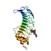
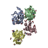
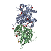
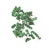
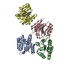
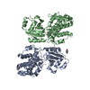
- PDB-6xfq: Structure of a novel antithrombotic agent Agkisacucetin in comple... -
+ Open data

- Basic information
| Entry | Database: PDB / ID: 6xfq | ||||||
|---|---|---|---|---|---|---|---|
| Title | Structure of a novel antithrombotic agent Agkisacucetin in complex with the platelet glycoprotein Ib receptor | ||||||
 Components Components |
| ||||||
 Keywords Keywords | STRUCTURAL PROTEIN / complex / GPIb | ||||||
| Function / homology |  Function and homology information Function and homology informationthrombin-activated receptor activity / glycoprotein Ib-IX-V complex / Enhanced binding of GP1BA variant to VWF multimer:collagen / Defective binding of VWF variant to GPIb:IX:V / blood coagulation, intrinsic pathway / Defective F9 activation / Platelet Adhesion to exposed collagen / positive regulation of platelet activation / megakaryocyte development / GP1b-IX-V activation signalling ...thrombin-activated receptor activity / glycoprotein Ib-IX-V complex / Enhanced binding of GP1BA variant to VWF multimer:collagen / Defective binding of VWF variant to GPIb:IX:V / blood coagulation, intrinsic pathway / Defective F9 activation / Platelet Adhesion to exposed collagen / positive regulation of platelet activation / megakaryocyte development / GP1b-IX-V activation signalling / regulation of blood coagulation / Platelet Aggregation (Plug Formation) / fibrinolysis / Intrinsic Pathway of Fibrin Clot Formation / release of sequestered calcium ion into cytosol / RUNX1 regulates genes involved in megakaryocyte differentiation and platelet function / platelet activation / extracellular matrix / cell morphogenesis / blood coagulation / toxin activity / cell surface receptor signaling pathway / cell adhesion / external side of plasma membrane / cell surface / extracellular space / extracellular exosome / extracellular region / membrane / plasma membrane Similarity search - Function | ||||||
| Biological species |  Homo sapiens (human) Homo sapiens (human) Deinagkistrodon acutus (Chinese moccasin) Deinagkistrodon acutus (Chinese moccasin) | ||||||
| Method |  X-RAY DIFFRACTION / SYNCHROTRON / MOLECULAR REPLACEMENT / molecular replacement / Resolution: 3.3 Å X-RAY DIFFRACTION / SYNCHROTRON / MOLECULAR REPLACEMENT / molecular replacement / Resolution: 3.3 Å | ||||||
 Authors Authors | Wang, J. / Gao, Y.X. / Ke, J.Y. / Zhu, Z.L. / Niu, L.W. | ||||||
| Funding support |  China, 1items China, 1items
| ||||||
 Citation Citation | Journal: To be published Title: Structure of a novel antithrombotic agent Agkisacucetin in complex with the platelet glycoprotein Ib receptor Authors: Wang, J. / Gao, Y.X. / Ke, J.Y. / Zhu, Z.L. / Niu, L.W. | ||||||
| History |
|
- Structure visualization
| Structure viewer | Molecule: MolmilJmol/JSmol |
|---|
- Downloads & links
-Download
| PDBx/mmCIF format | 6xfq.cif.gz | 120.4 KB | Display | PDBx/mmCIF format |
|---|---|---|---|---|
| PDB format | pdb6xfq.ent.gz | 89.6 KB | Display | PDB format |
| PDBx/mmJSON format | 6xfq.json.gz | Tree view | PDBx/mmJSON format | |
| Others |  Other downloads Other downloads |
-Validation report
| Arichive directory | https://data.pdbj.org/pub/pdb/validation_reports/xf/6xfqftp://data.pdbj.org/pub/pdb/validation_reports/xf/6xfq | HTTPS FTP |
|---|
-Related structure data


-Links
 PDBj
PDBj


- Assembly
| Deposited unit | 
| ||||||||||
|---|---|---|---|---|---|---|---|---|---|---|---|
| 1 |
| ||||||||||
| Unit cell |
|
-Components
| #1: Protein | Mass: 34010.820 Da / Num. of mol.: 1 Source method: isolated from a genetically manipulated source Source: (gene. exp.) Homo sapiens (human) / Gene: GP1BA / Production host:   Cricetulus griseus (Chinese hamster) / Strain (production host): CHO-DE44 / References: UniProt: P07359 Cricetulus griseus (Chinese hamster) / Strain (production host): CHO-DE44 / References: UniProt: P07359 |
|---|---|
| #2: Protein | Mass: 17334.500 Da / Num. of mol.: 1 Source method: isolated from a genetically manipulated source Source: (gene. exp.) Deinagkistrodon acutus (Chinese moccasin)Production host:  Komagataella pastoris (fungus) / References: UniProt: Q8JIV9 Komagataella pastoris (fungus) / References: UniProt: Q8JIV9 |
| #3: Protein | Mass: 17258.977 Da / Num. of mol.: 1 Source method: isolated from a genetically manipulated source Source: (gene. exp.) Deinagkistrodon acutus (Chinese moccasin)Production host: Komagataella pastoris (fungus) / References: UniProt: Q8AYA3 |
| #4: Water | ChemComp-HOH /  Mass: 18.015 Da / Num. of mol.: 6 / Source method: isolated from a natural source / Formula: H2O Mass: 18.015 Da / Num. of mol.: 6 / Source method: isolated from a natural source / Formula: H2O |
| Has protein modification | Y |
-Experimental details
-Experiment
| Experiment | Method: X-RAY DIFFRACTION / Number of used crystals: 1 |
|---|
- Sample preparation
| Crystal | Density Matthews: 2.58 Å3/Da / Density % sol: 52.29 % |
|---|---|
| Crystal grow | Temperature: 289 K / Method: vapor diffusion, hanging drop / Details: 15% PEG4000, 0.1M HEPES, pH 7.5 |
-Data collection
| Diffraction | Mean temperature: 100 K / Serial crystal experiment: N |
|---|---|
| Diffraction source | Source: SYNCHROTRON / Site: SSRF / Beamline: BL17U / Wavelength: 0.97915 Å |
| Detector | Type: ADSC QUANTUM 315r / Detector: CCD / Date: Apr 13, 2014 |
| Radiation | Protocol: SINGLE WAVELENGTH / Monochromatic (M) / Laue (L): M / Scattering type: x-ray |
| Radiation wavelength | Wavelength: 0.97915 Å / Relative weight: 1 |
| Reflection | Resolution: 3.3→58.1 Å / Num. obs: 11456 / % possible obs: 99.9 % / Redundancy: 8.2 % / Biso Wilson estimate: 115.46 Å2 / CC1/2: 0.99 / Rmerge(I) obs: 0.154 / Rpim(I) all: 0.057 / Net I/σ(I): 9.8 |
| Reflection shell | Resolution: 3.3→3.56 Å / Redundancy: 8.6 % / Num. unique obs: 2290 / CC1/2: 0.918 / Rpim(I) all: 0.674 / % possible all: 99.9 |
-Phasing
| Phasing | Method: molecular replacement |
|---|
- Processing
| Software |
| |||||||||||||||||||||||||||||||||||
|---|---|---|---|---|---|---|---|---|---|---|---|---|---|---|---|---|---|---|---|---|---|---|---|---|---|---|---|---|---|---|---|---|---|---|---|---|
| Refinement | Method to determine structure: MOLECULAR REPLACEMENT Starting model: 1P9A, 3UBU Resolution: 3.3→51.34 Å / SU ML: 0.546 / Cross valid method: THROUGHOUT / σ(F): 1.33 / Phase error: 44.5898 / Stereochemistry target values: CDL v1.2
| |||||||||||||||||||||||||||||||||||
| Solvent computation | Shrinkage radii: 0.9 Å / VDW probe radii: 1.11 Å / Solvent model: FLAT BULK SOLVENT MODEL | |||||||||||||||||||||||||||||||||||
| Displacement parameters | Biso mean: 162.83 Å2 | |||||||||||||||||||||||||||||||||||
| Refinement step | Cycle: LAST / Resolution: 3.3→51.34 Å
| |||||||||||||||||||||||||||||||||||
| Refine LS restraints |
| |||||||||||||||||||||||||||||||||||
| LS refinement shell |
|
