 Movie
Movie Controller


+ Open data

- Basic information
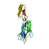
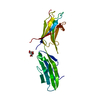
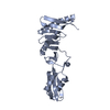
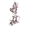
| Entry | Database: PDB / ID: 6x3k | ||||||||||||
|---|---|---|---|---|---|---|---|---|---|---|---|---|---|
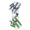
| Title | Hsa Siglec and Unique domains in complex with 6S-sialy-Lewisx | ||||||||||||
 Components Components | Streptococcal hemagglutinin | ||||||||||||
 Keywords Keywords | CELL ADHESION / adhesin / serine rich repeat adhesin / bacterial adhesion / protein | ||||||||||||
| Function / homology |  Function and homology information Function and homology information | ||||||||||||
| Biological species |  Streptococcus gordonii str. Challis (bacteria) Streptococcus gordonii str. Challis (bacteria) | ||||||||||||
| Method |  X-RAY DIFFRACTION / SYNCHROTRON / MOLECULAR REPLACEMENT / Resolution: 2.5 Å X-RAY DIFFRACTION / SYNCHROTRON / MOLECULAR REPLACEMENT / Resolution: 2.5 Å | ||||||||||||
 Authors Authors | Stubbs, H.E. / Iverson, T.M. | ||||||||||||
| Funding support | 3items
| ||||||||||||
 Citation Citation | Journal: Nat Commun / Year: 2022 Title: Origins of glycan selectivity in streptococcal Siglec-like adhesins suggest mechanisms of receptor adaptation. Authors: Bensing, B.A. / Stubbs, H.E. / Agarwal, R. / Yamakawa, I. / Luong, K. / Solakyildirim, K. / Yu, H. / Hadadianpour, A. / Castro, M.A. / Fialkowski, K.P. / Morrison, K.M. / Wawrzak, Z. / Chen, ...Authors: Bensing, B.A. / Stubbs, H.E. / Agarwal, R. / Yamakawa, I. / Luong, K. / Solakyildirim, K. / Yu, H. / Hadadianpour, A. / Castro, M.A. / Fialkowski, K.P. / Morrison, K.M. / Wawrzak, Z. / Chen, X. / Lebrilla, C.B. / Baudry, J. / Smith, J.C. / Sullam, P.M. / Iverson, T.M. | ||||||||||||
| History |
|
- Structure visualization
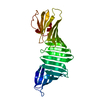
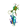
| Structure viewer | Molecule: MolmilJmol/JSmol |
|---|
- Downloads & links
-Download
| PDBx/mmCIF format | 6x3k.cif.gz | 103.4 KB | Display | PDBx/mmCIF format |
|---|---|---|---|---|
| PDB format | pdb6x3k.ent.gz | 72.4 KB | Display | PDB format |
| PDBx/mmJSON format | 6x3k.json.gz | Tree view | PDBx/mmJSON format | |
| Others |  Other downloads Other downloads |
-Validation report
| Arichive directory | https://data.pdbj.org/pub/pdb/validation_reports/x3/6x3kftp://data.pdbj.org/pub/pdb/validation_reports/x3/6x3k | HTTPS FTP |
|---|
-Related structure data
| Related structure data |  6ef7C  6ef9C  6efaC  6efbC  6efcSC 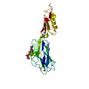 6efdC  6effC  6efiC 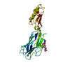 6x3qC  7kmjC S: Starting model for refinement C: citing same article ( |
|---|---|
| Similar structure data |

-Links
 PDBj
PDBj

- Assembly
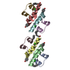
| Deposited unit | 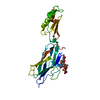
| ||||||||||||
|---|---|---|---|---|---|---|---|---|---|---|---|---|---|
| 1 |
| ||||||||||||
| Unit cell |
|
-Components
| #1: Protein | Mass: 25837.381 Da / Num. of mol.: 1 Source method: isolated from a genetically manipulated source Source: (gene. exp.) Streptococcus gordonii str. Challis (bacteria)Gene: hsa / Production host: | ||||||
|---|---|---|---|---|---|---|---|
| #2: Polysaccharide | alpha-L-fucopyranose-(1-3)-[beta-D-galactopyranose-(1-4)]2-acetamido-2-deoxy-6-O-sulfo-beta-D-glucopyranose Source method: isolated from a genetically manipulated source | ||||||
| #3: Chemical |   Mass: 22.990 Da / Num. of mol.: 3 / Source method: obtained synthetically / Formula: Na Mass: 22.990 Da / Num. of mol.: 3 / Source method: obtained synthetically / Formula: Na#4: Sugar | ChemComp-SIA / |   Type: D-saccharide, alpha linking / Mass: 309.270 Da / Num. of mol.: 1 Type: D-saccharide, alpha linking / Mass: 309.270 Da / Num. of mol.: 1Source method: isolated from a genetically manipulated source Formula: C11H19NO9 / Feature type: SUBJECT OF INVESTIGATION #5: Water | ChemComp-HOH / |  Mass: 18.015 Da / Num. of mol.: 17 / Source method: isolated from a natural source / Formula: H2O Mass: 18.015 Da / Num. of mol.: 17 / Source method: isolated from a natural source / Formula: H2OHas ligand of interest | Y | |
-Experimental details
-Experiment
| Experiment | Method: X-RAY DIFFRACTION / Number of used crystals: 1 |
|---|
- Sample preparation
| Crystal | Density Matthews: 1.99 Å3/Da / Density % sol: 38.32 % |
|---|---|
| Crystal grow | Temperature: 298 K / Method: vapor diffusion, sitting drop Details: 21.6 mg/ml protein in 20 mM Tris-HCl, pH 7.2, Mix 1uL protein solution with 2uL reservoir solution. Reservoir solution: 0.1 M Succinate/Phosphate/Glycine pH 10.0 and 25% PEG 3350 |
-Data collection
| Diffraction | Mean temperature: 100 K / Serial crystal experiment: N |
|---|---|
| Diffraction source | Source: SYNCHROTRON / Site: SSRL  / Beamline: BL9-2 / Wavelength: 0.979 Å / Beamline: BL9-2 / Wavelength: 0.979 Å |
| Detector | Type: DECTRIS PILATUS 6M / Detector: PIXEL / Date: Nov 21, 2019 |
| Radiation | Protocol: SINGLE WAVELENGTH / Monochromatic (M) / Laue (L): M / Scattering type: x-ray |
| Radiation wavelength | Wavelength: 0.979 Å / Relative weight: 1 |
| Reflection | Resolution: 2.5→50 Å / Num. obs: 7619 / % possible obs: 98.7 % / Redundancy: 4.9 % / Biso Wilson estimate: 49.17 Å2 / CC1/2: 0.989 / Net I/σ(I): 15.6 |
| Reflection shell | Resolution: 2.5→2.54 Å / Num. unique obs: 363 / Rpim(I) all: 0.318 / Rsym value: 0.74 |
- Processing
| Software |
| ||||||||||||||||||||||||||||
|---|---|---|---|---|---|---|---|---|---|---|---|---|---|---|---|---|---|---|---|---|---|---|---|---|---|---|---|---|---|
| Refinement | Method to determine structure: MOLECULAR REPLACEMENT Starting model: 6efc Resolution: 2.5→50 Å / SU ML: 0.3411 / Cross valid method: FREE R-VALUE / σ(F): 1.36 / Phase error: 29.0637 Stereochemistry target values: GeoStd + Monomer Library + CDL v1.2
| ||||||||||||||||||||||||||||
| Solvent computation | Shrinkage radii: 0.9 Å / VDW probe radii: 1.11 Å / Solvent model: FLAT BULK SOLVENT MODEL | ||||||||||||||||||||||||||||
| Displacement parameters | Biso mean: 55.06 Å2 | ||||||||||||||||||||||||||||
| Refinement step | Cycle: LAST / Resolution: 2.5→50 Å
| ||||||||||||||||||||||||||||
| Refine LS restraints |
| ||||||||||||||||||||||||||||
| LS refinement shell |
|
