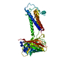
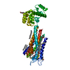
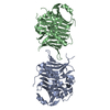
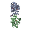
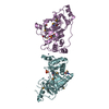
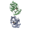
Green fluorescent protein, Protein jagunal homolog 1 chimera
Keywords
MEMBRANE PROTEIN / JAGN1 / Protein jagunal homolog 1
Function / homology
Function and homology information
granulocyte colony-stimulating factor signaling pathway / neutrophil differentiation / negative regulation of insulin secretion involved in cellular response to glucose stimulus / neutrophil migration / endoplasmic reticulum organization / neutrophil mediated immunity / exocytosis / defense response to fungus / vesicle-mediated transport / bioluminescence ...granulocyte colony-stimulating factor signaling pathway / neutrophil differentiation / negative regulation of insulin secretion involved in cellular response to glucose stimulus / neutrophil migration / endoplasmic reticulum organization / neutrophil mediated immunity / exocytosis / defense response to fungus / vesicle-mediated transport / bioluminescence / generation of precursor metabolites and energy / protein transport / endoplasmic reticulum membrane / endoplasmic reticulum Similarity search - Function
Protein jagunal / Jagunal, ER re-organisation during oogenesis / Green fluorescent protein, GFP / Green fluorescent protein-related / Green fluorescent protein / Green fluorescent protein Similarity search - Domain/homology
(2R)-2,3-dihydroxypropyl (9Z)-octadec-9-enoate / Green fluorescent protein / Protein jagunal homolog 1 Similarity search - Component
Biological species
Aequorea victoria (jellyfish) Homo sapiens (human)
In the structure databanks used in Yorodumi, some data are registered as the other names, "COVID-19 virus" and "2019-nCoV". Here are the details of the virus and the list of structure data.
Jan 31, 2019. EMDB accession codes are about to change! (news from PDBe EMDB page)
EMDB accession codes are about to change! (news from PDBe EMDB page)
The allocation of 4 digits for EMDB accession codes will soon come to an end. Whilst these codes will remain in use, new EMDB accession codes will include an additional digit and will expand incrementally as the available range of codes is exhausted. The current 4-digit format prefixed with “EMD-” (i.e. EMD-XXXX) will advance to a 5-digit format (i.e. EMD-XXXXX), and so on. It is currently estimated that the 4-digit codes will be depleted around Spring 2019, at which point the 5-digit format will come into force.
The EM Navigator/Yorodumi systems omit the EMD- prefix.
Related info.:Q: What is EMD? / ID/Accession-code notation in Yorodumi/EM Navigator
Yorodumi is a browser for structure data from EMDB, PDB, SASBDB, etc.
This page is also the successor to EM Navigator detail page, and also detail information page/front-end page for Omokage search.
The word "yorodu" (or yorozu) is an old Japanese word meaning "ten thousand". "mi" (miru) is to see.
Related info.:EMDB / PDB / SASBDB / Comparison of 3 databanks / Yorodumi Search / Aug 31, 2016. New EM Navigator & Yorodumi / Yorodumi Papers / Jmol/JSmol / Function and homology information / Changes in new EM Navigator and Yorodumi
 Movie
Movie Controller


 Open data
 Basic information
 Components
 Keywords
 Function and homology information
 Aequorea victoria (jellyfish)
 Homo sapiens (human)
 X-RAY DIFFRACTION /
  Authors
 United States, 6items
  Citation
 Structure visualization
 Downloads & links
 Other downloads


 PDBj
PDBj


 Assembly
 Komagataella pastoris (fungus) / References: UniProt: P42212, UniProt: Q8N5M9
 Mass: 356.540 Da / Num. of mol.: 2 / Source method: obtained synthetically / Formula: C21H40O4
 Mass: 18.015 Da / Num. of mol.: 95 / Source method: isolated from a natural source / Formula: H2O
 Sample preparation
 Processing