 Movie
Movie Controller


+ Open data

- Basic information
| Entry | Database: PDB / ID: 6wv6 | |||||||||||||||||||||
|---|---|---|---|---|---|---|---|---|---|---|---|---|---|---|---|---|---|---|---|---|---|---|
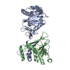
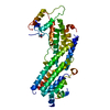
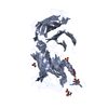
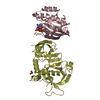
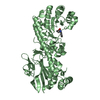
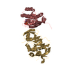
| Title | Human VKOR with phenindione | |||||||||||||||||||||
 Components Components | Vitamin K epoxide reductase, termini restrained by green fluorescent protein | |||||||||||||||||||||
 Keywords Keywords | OXIDOREDUCTASE / FLUORESCENT PROTEIN / Vitamin K epoxide Reductase (VKOR) / Vitamin K / warfarin / superwarfarin / phenindione / vitamin K expoxide(KO) / membrane protein | |||||||||||||||||||||
| Function / homology |  Function and homology information Function and homology informationpeptidyl-glutamic acid carboxylation / vitamin-K-epoxide reductase (warfarin-insensitive) activity / Metabolism of vitamin K / vitamin-K-epoxide reductase (warfarin-sensitive) / vitamin-K-epoxide reductase (warfarin-sensitive) activity / positive regulation of coagulation / vitamin K metabolic process / quinone binding / xenobiotic metabolic process / bioluminescence ...peptidyl-glutamic acid carboxylation / vitamin-K-epoxide reductase (warfarin-insensitive) activity / Metabolism of vitamin K / vitamin-K-epoxide reductase (warfarin-sensitive) / vitamin-K-epoxide reductase (warfarin-sensitive) activity / positive regulation of coagulation / vitamin K metabolic process / quinone binding / xenobiotic metabolic process / bioluminescence / generation of precursor metabolites and energy / bone development / blood coagulation / endoplasmic reticulum lumen / endoplasmic reticulum membrane / endoplasmic reticulum Similarity search - Function | |||||||||||||||||||||
| Biological species |   Aequorea victoria (jellyfish) Aequorea victoria (jellyfish) Homo sapiens (human) Homo sapiens (human) | |||||||||||||||||||||
| Method |  X-RAY DIFFRACTION / SYNCHROTRON / MOLECULAR REPLACEMENT / Resolution: 2.7 Å X-RAY DIFFRACTION / SYNCHROTRON / MOLECULAR REPLACEMENT / Resolution: 2.7 Å | |||||||||||||||||||||
 Authors Authors | Liu, S. / Sukumar, N. / Li, W. | |||||||||||||||||||||
| Funding support |  United States, 6items United States, 6items
| |||||||||||||||||||||
 Citation Citation | Journal: Science / Year: 2021 Title: Structural basis of antagonizing the vitamin K catalytic cycle for anticoagulation. Authors: Liu, S. / Li, S. / Shen, G. / Sukumar, N. / Krezel, A.M. / Li, W. #1: Journal: To Be PublishedTitle: Termini restraining of small membrane proteins enables structure determination at atomic resolution Authors: Liu, S. / Li, S. / Yang, Y. / Li, W. | |||||||||||||||||||||
| History |
|
- Structure visualization
| Structure viewer | Molecule: MolmilJmol/JSmol |
|---|
- Downloads & links
-Download
| PDBx/mmCIF format | 6wv6.cif.gz | 171.1 KB | Display | PDBx/mmCIF format |
|---|---|---|---|---|
| PDB format | pdb6wv6.ent.gz | 133.1 KB | Display | PDB format |
| PDBx/mmJSON format | 6wv6.json.gz | Tree view | PDBx/mmJSON format | |
| Others |  Other downloads Other downloads |
-Validation report
| Arichive directory | https://data.pdbj.org/pub/pdb/validation_reports/wv/6wv6ftp://data.pdbj.org/pub/pdb/validation_reports/wv/6wv6 | HTTPS FTP |
|---|
-Related structure data
| Related structure data |  6wv3C  6wv4C  6wv5C  6wv7C  6wv8C 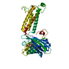 6wv9C 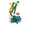 6wvaC  6wvbC  6wvhC  6wviC  2b3pS S: Starting model for refinement C: citing same article ( |
|---|---|
| Similar structure data |


-Links
 PDBj
PDBj


- Assembly
| Deposited unit | 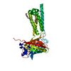
| ||||||||
|---|---|---|---|---|---|---|---|---|---|
| 1 |
| ||||||||
| Unit cell |
|
-Components
| #1: Protein | Mass: 44197.434 Da / Num. of mol.: 1 Fragment: GPF (UNP residues 1-144) + VKOR + GFP (UNP residues 146-231) Source method: isolated from a genetically manipulated source Source: (gene. exp.) Aequorea victoria (jellyfish), (gene. exp.) Homo sapiens (human)Gene: gfp, VKORC1, VKOR, MSTP134, MSTP576, UNQ308/PRO351 / Production host:  Komagataella pastoris (fungus) Komagataella pastoris (fungus)References: UniProt: A0A059PIQ0, UniProt: Q9BQB6, UniProt: P42212*PLUS, vitamin-K-epoxide reductase (warfarin-sensitive) | ||||||||
|---|---|---|---|---|---|---|---|---|---|
| #2: Chemical |   Mass: 356.540 Da / Num. of mol.: 2 / Source method: obtained synthetically / Formula: C21H40O4 Mass: 356.540 Da / Num. of mol.: 2 / Source method: obtained synthetically / Formula: C21H40O4#3: Chemical | ChemComp-UAS / |   Mass: 222.239 Da / Num. of mol.: 1 / Source method: obtained synthetically / Formula: C15H10O2 / Feature type: SUBJECT OF INVESTIGATION / Comment: anticoagulant, antagonist*YM Mass: 222.239 Da / Num. of mol.: 1 / Source method: obtained synthetically / Formula: C15H10O2 / Feature type: SUBJECT OF INVESTIGATION / Comment: anticoagulant, antagonist*YM#4: Water | ChemComp-HOH / |  Mass: 18.015 Da / Num. of mol.: 33 / Source method: isolated from a natural source / Formula: H2O Mass: 18.015 Da / Num. of mol.: 33 / Source method: isolated from a natural source / Formula: H2OHas ligand of interest | Y | Has protein modification | Y | |
-Experimental details
-Experiment
| Experiment | Method: X-RAY DIFFRACTION / Number of used crystals: 1 |
|---|
- Sample preparation
| Crystal | Density Matthews: 3.17 Å3/Da / Density % sol: 61.17 % |
|---|---|
| Crystal grow | Temperature: 295 K / Method: lipidic cubic phase / pH: 6.5 Details: 34% PEG400, 80 mM lithium sulfate, 0.1 M MES, pH 6.5 |
-Data collection
| Diffraction | Mean temperature: 100 K / Serial crystal experiment: N | |||||||||||||||||||||||||||||||||||||||||||||||||||||||||||||||||||||||||||||||||||||||||||||||||||||||||||||||||||||||||||||||||||||||||||||||||||||||||||||||||||||||||||||||||||||||||||||
|---|---|---|---|---|---|---|---|---|---|---|---|---|---|---|---|---|---|---|---|---|---|---|---|---|---|---|---|---|---|---|---|---|---|---|---|---|---|---|---|---|---|---|---|---|---|---|---|---|---|---|---|---|---|---|---|---|---|---|---|---|---|---|---|---|---|---|---|---|---|---|---|---|---|---|---|---|---|---|---|---|---|---|---|---|---|---|---|---|---|---|---|---|---|---|---|---|---|---|---|---|---|---|---|---|---|---|---|---|---|---|---|---|---|---|---|---|---|---|---|---|---|---|---|---|---|---|---|---|---|---|---|---|---|---|---|---|---|---|---|---|---|---|---|---|---|---|---|---|---|---|---|---|---|---|---|---|---|---|---|---|---|---|---|---|---|---|---|---|---|---|---|---|---|---|---|---|---|---|---|---|---|---|---|---|---|---|---|---|---|---|
| Diffraction source | Source: SYNCHROTRON / Site: APS / Beamline: 24-ID-C / Wavelength: 0.9791 Å | |||||||||||||||||||||||||||||||||||||||||||||||||||||||||||||||||||||||||||||||||||||||||||||||||||||||||||||||||||||||||||||||||||||||||||||||||||||||||||||||||||||||||||||||||||||||||||||
| Detector | Type: DECTRIS PILATUS 6M-F / Detector: PIXEL / Date: Dec 6, 2016 | |||||||||||||||||||||||||||||||||||||||||||||||||||||||||||||||||||||||||||||||||||||||||||||||||||||||||||||||||||||||||||||||||||||||||||||||||||||||||||||||||||||||||||||||||||||||||||||
| Radiation | Monochromator: double crystal Si(111) / Protocol: SINGLE WAVELENGTH / Monochromatic (M) / Laue (L): M / Scattering type: x-ray | |||||||||||||||||||||||||||||||||||||||||||||||||||||||||||||||||||||||||||||||||||||||||||||||||||||||||||||||||||||||||||||||||||||||||||||||||||||||||||||||||||||||||||||||||||||||||||||
| Radiation wavelength | Wavelength: 0.9791 Å / Relative weight: 1 | |||||||||||||||||||||||||||||||||||||||||||||||||||||||||||||||||||||||||||||||||||||||||||||||||||||||||||||||||||||||||||||||||||||||||||||||||||||||||||||||||||||||||||||||||||||||||||||
| Reflection | Resolution: 2.7→50 Å / Num. obs: 15647 / % possible obs: 97.1 % / Redundancy: 2.9 % / Rmerge(I) obs: 0.117 / Rpim(I) all: 0.079 / Rrim(I) all: 0.142 / Χ2: 1.156 / Net I/σ(I): 6.8 / Num. measured all: 45677 | |||||||||||||||||||||||||||||||||||||||||||||||||||||||||||||||||||||||||||||||||||||||||||||||||||||||||||||||||||||||||||||||||||||||||||||||||||||||||||||||||||||||||||||||||||||||||||||
| Reflection shell | Diffraction-ID: 1
|
- Processing
| Software |
| |||||||||||||||||||||||||||||||||||||||||||||||||||||||||||||||||||||||||||
|---|---|---|---|---|---|---|---|---|---|---|---|---|---|---|---|---|---|---|---|---|---|---|---|---|---|---|---|---|---|---|---|---|---|---|---|---|---|---|---|---|---|---|---|---|---|---|---|---|---|---|---|---|---|---|---|---|---|---|---|---|---|---|---|---|---|---|---|---|---|---|---|---|---|---|---|---|
| Refinement | Method to determine structure: MOLECULAR REPLACEMENT Starting model: PDB entry 2B3P Resolution: 2.7→37.3 Å / SU ML: 0.43 / Cross valid method: THROUGHOUT / σ(F): 1.35 / Phase error: 28.11 / Stereochemistry target values: ML
| |||||||||||||||||||||||||||||||||||||||||||||||||||||||||||||||||||||||||||
| Solvent computation | Shrinkage radii: 0.9 Å / VDW probe radii: 1.11 Å / Solvent model: FLAT BULK SOLVENT MODEL | |||||||||||||||||||||||||||||||||||||||||||||||||||||||||||||||||||||||||||
| Displacement parameters | Biso max: 114.43 Å2 / Biso mean: 50.9532 Å2 / Biso min: 17.95 Å2 | |||||||||||||||||||||||||||||||||||||||||||||||||||||||||||||||||||||||||||
| Refinement step | Cycle: final / Resolution: 2.7→37.3 Å
| |||||||||||||||||||||||||||||||||||||||||||||||||||||||||||||||||||||||||||
| LS refinement shell | Refine-ID: X-RAY DIFFRACTION / Rfactor Rfree error: 0 / Total num. of bins used: 5
| |||||||||||||||||||||||||||||||||||||||||||||||||||||||||||||||||||||||||||
| Refinement TLS params. | Method: refined / Refine-ID: X-RAY DIFFRACTION
| |||||||||||||||||||||||||||||||||||||||||||||||||||||||||||||||||||||||||||
| Refinement TLS group |
|
