 Movie
Movie Controller

[English] 日本語
 Yorodumi
- PDB-6wrk: Crystal structure of 3rd-generation Mj 3-nitro-tyrosine tRNA synt... -
+ Open data

- Basic information
| Entry | Database: PDB / ID: 6wrk | ||||||||||||
|---|---|---|---|---|---|---|---|---|---|---|---|---|---|
| Title | Crystal structure of 3rd-generation Mj 3-nitro-tyrosine tRNA synthetase ("A7") bound to 3-nitro-tyrosine | ||||||||||||
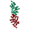
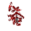
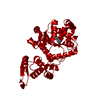
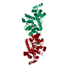
 Components Components | Tyrosine--tRNA ligase | ||||||||||||
 Keywords Keywords | LIGASE / aminoacyl-tRNA synthetase / 3-nitro-tyrosine | ||||||||||||
| Function / homology |  Function and homology information Function and homology informationtyrosyl-tRNA aminoacylation / tyrosine-tRNA ligase / tyrosine-tRNA ligase activity / ATP binding / cytoplasm Similarity search - Function | ||||||||||||
| Biological species |   Methanocaldococcus jannaschii (archaea) Methanocaldococcus jannaschii (archaea) | ||||||||||||
| Method |  X-RAY DIFFRACTION / SYNCHROTRON / MOLECULAR REPLACEMENT / Resolution: 1.95 Å X-RAY DIFFRACTION / SYNCHROTRON / MOLECULAR REPLACEMENT / Resolution: 1.95 Å | ||||||||||||
 Authors Authors | Beyer, J.N. / Hosseinzadeh, P. / Karplus, P.A. / Mehl, R.A. / Cooley, R.B. | ||||||||||||
| Funding support |  United States, 3items United States, 3items
| ||||||||||||
 Citation Citation | Journal: J.Mol.Biol. / Year: 2020 Title: Overcoming Near-Cognate Suppression in a Release Factor 1-Deficient Host with an Improved Nitro-Tyrosine tRNA Synthetase. Authors: Beyer, J.N. / Hosseinzadeh, P. / Gottfried-Lee, I. / Van Fossen, E.M. / Zhu, P. / Bednar, R.M. / Karplus, P.A. / Mehl, R.A. / Cooley, R.B. | ||||||||||||
| History |
|
- Structure visualization
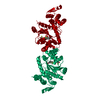
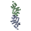
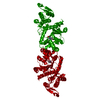
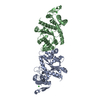
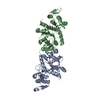
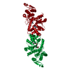
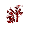
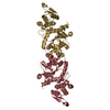
| Structure viewer | Molecule: MolmilJmol/JSmol |
|---|
- Downloads & links
-Download
| PDBx/mmCIF format | 6wrk.cif.gz | 146.7 KB | Display | PDBx/mmCIF format |
|---|---|---|---|---|
| PDB format | pdb6wrk.ent.gz | 115.2 KB | Display | PDB format |
| PDBx/mmJSON format | 6wrk.json.gz | Tree view | PDBx/mmJSON format | |
| Others |  Other downloads Other downloads |
-Validation report
| Arichive directory | https://data.pdbj.org/pub/pdb/validation_reports/wr/6wrkftp://data.pdbj.org/pub/pdb/validation_reports/wr/6wrk | HTTPS FTP |
|---|
-Related structure data
| Related structure data | 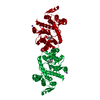 6wrnC 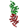 6wrqC 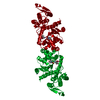 6wrtC 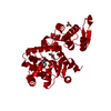 4ndaS C: citing same article ( S: Starting model for refinement |
|---|---|
| Similar structure data |


-Links
 PDBj
PDBj
- Assembly
| Deposited unit | 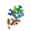
| ||||||||||||
|---|---|---|---|---|---|---|---|---|---|---|---|---|---|
| 1 | 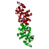
| ||||||||||||
| Unit cell |
| ||||||||||||
| Components on special symmetry positions |
|
-Components
| #1: Protein | Mass: 36136.898 Da / Num. of mol.: 1 / Mutation: Y32H, H70T, D158H, I159A, L162R Source method: isolated from a genetically manipulated source Source: (gene. exp.) Methanocaldococcus jannaschii (strain ATCC 43067 / DSM 2661 / JAL-1 / JCM 10045 / NBRC 100440) (archaea)Strain: ATCC 43067 / DSM 2661 / JAL-1 / JCM 10045 / NBRC 100440 Gene: tyrS, MJ0389 / Production host:  | ||||||||
|---|---|---|---|---|---|---|---|---|---|
| #2: Chemical |   Mass: 92.094 Da / Num. of mol.: 3 / Source method: obtained synthetically / Formula: C3H8O3 Mass: 92.094 Da / Num. of mol.: 3 / Source method: obtained synthetically / Formula: C3H8O3#3: Chemical | ChemComp-NA / |   Mass: 22.990 Da / Num. of mol.: 1 / Source method: obtained synthetically / Formula: Na Mass: 22.990 Da / Num. of mol.: 1 / Source method: obtained synthetically / Formula: Na#4: Chemical | 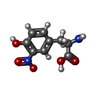  Type: L-peptide linking / Mass: 226.186 Da / Num. of mol.: 2 / Source method: obtained synthetically / Formula: C9H10N2O5 / Feature type: SUBJECT OF INVESTIGATION Type: L-peptide linking / Mass: 226.186 Da / Num. of mol.: 2 / Source method: obtained synthetically / Formula: C9H10N2O5 / Feature type: SUBJECT OF INVESTIGATION#5: Water | ChemComp-HOH / |  Mass: 18.015 Da / Num. of mol.: 155 / Source method: isolated from a natural source / Formula: H2O Mass: 18.015 Da / Num. of mol.: 155 / Source method: isolated from a natural source / Formula: H2OHas ligand of interest | Y | |
-Experimental details
-Experiment
| Experiment | Method: X-RAY DIFFRACTION / Number of used crystals: 1 |
|---|
- Sample preparation
| Crystal | Density Matthews: 2.54 Å3/Da / Density % sol: 51.66 % |
|---|---|
| Crystal grow | Temperature: 298 K / Method: vapor diffusion, hanging drop Details: 22-23% PEG 300, 5% PEG 8000, 10% glycerol and 100 mM Tris pH 7.9 to 8.2 PH range: 7.9-8.2 |
-Data collection
| Diffraction | Mean temperature: 100 K / Serial crystal experiment: N |
|---|---|
| Diffraction source | Source: SYNCHROTRON / Site: ALS / Beamline: 5.0.2 / Wavelength: 1 Å |
| Detector | Type: DECTRIS PILATUS3 6M / Detector: PIXEL / Date: Jun 22, 2019 |
| Radiation | Monochromator: Si / Protocol: SINGLE WAVELENGTH / Monochromatic (M) / Laue (L): M / Scattering type: x-ray |
| Radiation wavelength | Wavelength: 1 Å / Relative weight: 1 |
| Reflection | Resolution: 1.95→45.36 Å / Num. obs: 28169 / % possible obs: 100 % / Redundancy: 13.8 % / CC1/2: 0.998 / Net I/σ(I): 10.17 |
| Reflection shell | Resolution: 1.95→2.05 Å / Redundancy: 13.8 % / Mean I/σ(I) obs: 0.9 / Num. unique obs: 7238 / CC1/2: 0.31 / % possible all: 100 |
- Processing
| Software |
| |||||||||||||||||||||||||||||||||||||||||||||||||
|---|---|---|---|---|---|---|---|---|---|---|---|---|---|---|---|---|---|---|---|---|---|---|---|---|---|---|---|---|---|---|---|---|---|---|---|---|---|---|---|---|---|---|---|---|---|---|---|---|---|---|
| Refinement | Method to determine structure: MOLECULAR REPLACEMENT Starting model: 4NDA Resolution: 1.95→45.36 Å / SU ML: 0.2811 / Cross valid method: FREE R-VALUE / σ(F): 1.36 / Phase error: 25.527 Stereochemistry target values: GeoStd + Monomer Library + CDL v1.2
| |||||||||||||||||||||||||||||||||||||||||||||||||
| Solvent computation | Shrinkage radii: 0.9 Å / VDW probe radii: 1.11 Å / Solvent model: FLAT BULK SOLVENT MODEL | |||||||||||||||||||||||||||||||||||||||||||||||||
| Displacement parameters | Biso mean: 43.99 Å2 | |||||||||||||||||||||||||||||||||||||||||||||||||
| Refinement step | Cycle: LAST / Resolution: 1.95→45.36 Å
| |||||||||||||||||||||||||||||||||||||||||||||||||
| Refine LS restraints |
| |||||||||||||||||||||||||||||||||||||||||||||||||
| LS refinement shell |
|
