 Movie
Movie Controller

[English] 日本語
 Yorodumi
- PDB-4pbs: Crystal structure of the M. jannaschii F9 tRNA synthetase variant... -
+ Open data

- Basic information
| Entry | Database: PDB / ID: 4pbs | ||||||
|---|---|---|---|---|---|---|---|
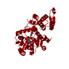
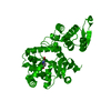
| Title | Crystal structure of the M. jannaschii F9 tRNA synthetase variant bound to 4-(2-bromoisobutyramido)-phenylalanine (BibaF) | ||||||
 Components Components | Tyrosine--tRNA ligase | ||||||
 Keywords Keywords | LIGASE / tRNA synthetase / non-natural amino acid | ||||||
| Function / homology |  Function and homology information Function and homology informationtyrosyl-tRNA aminoacylation / tyrosine-tRNA ligase / tyrosine-tRNA ligase activity / ATP binding / cytoplasm Similarity search - Function | ||||||
| Biological species |   Methanocaldococcus jannaschii (archaea) Methanocaldococcus jannaschii (archaea) | ||||||
| Method |  X-RAY DIFFRACTION / SYNCHROTRON / MOLECULAR REPLACEMENT / Resolution: 2.011 Å X-RAY DIFFRACTION / SYNCHROTRON / MOLECULAR REPLACEMENT / Resolution: 2.011 Å | ||||||
 Authors Authors | Cooley, R.B. / Karplus, P.A. / Mehl, R.A. | ||||||
| Funding support |  United States, 1items United States, 1items
| ||||||
 Citation Citation | Journal: Chembiochem / Year: 2014 Title: Gleaning Unexpected Fruits from Hard-Won Synthetases: Probing Principles of Permissivity in Non-canonical Amino Acid-tRNA Synthetases. Authors: Cooley, R.B. / Karplus, P.A. / Mehl, R.A. | ||||||
| History |
|
- Structure visualization
| Structure viewer | Molecule: MolmilJmol/JSmol |
|---|
- Downloads & links
-Download
| PDBx/mmCIF format | 4pbs.cif.gz | 145.8 KB | Display | PDBx/mmCIF format |
|---|---|---|---|---|
| PDB format | pdb4pbs.ent.gz | 114.1 KB | Display | PDB format |
| PDBx/mmJSON format | 4pbs.json.gz | Tree view | PDBx/mmJSON format | |
| Others |  Other downloads Other downloads |
-Validation report
| Arichive directory | https://data.pdbj.org/pub/pdb/validation_reports/pb/4pbsftp://data.pdbj.org/pub/pdb/validation_reports/pb/4pbs | HTTPS FTP |
|---|
-Related structure data
| Related structure data |  4pbrC  4pbtC  2zp1S C: citing same article ( S: Starting model for refinement |
|---|---|
| Similar structure data |

-Links
 PDBj
PDBj
- Assembly
| Deposited unit | 
| ||||||||
|---|---|---|---|---|---|---|---|---|---|
| 1 | 
| ||||||||
| Unit cell |
| ||||||||
| Components on special symmetry positions |
|
-Components
| #1: Protein | Mass: 36015.738 Da / Num. of mol.: 1 / Mutation: Y32G, L65E, D158G, I159C Source method: isolated from a genetically manipulated source Source: (gene. exp.) Methanocaldococcus jannaschii (archaea)Strain: ATCC 43067 / DSM 2661 / JAL-1 / JCM 10045 / NBRC 100440 Gene: tyrS, MJ0389 / Production host:  |
|---|---|
| #2: Chemical | ChemComp-2L7 /   Mass: 329.190 Da / Num. of mol.: 1 / Source method: obtained synthetically / Formula: C13H17BrN2O3 Mass: 329.190 Da / Num. of mol.: 1 / Source method: obtained synthetically / Formula: C13H17BrN2O3 |
| #3: Water | ChemComp-HOH /  Mass: 18.015 Da / Num. of mol.: 214 / Source method: isolated from a natural source / Formula: H2O Mass: 18.015 Da / Num. of mol.: 214 / Source method: isolated from a natural source / Formula: H2O |
-Experimental details
-Experiment
| Experiment | Method: X-RAY DIFFRACTION |
|---|
- Sample preparation
| Crystal | Density Matthews: 2.56 Å3/Da / Density % sol: 51.97 % |
|---|---|
| Crystal grow | Temperature: 293 K / Method: vapor diffusion, hanging drop / pH: 8 Details: 23% PEG 300, 5% PEG 8000, 10% glycerol and 100 mM Tris PH range: 7.8-8.2 |
-Data collection
| Diffraction | Mean temperature: 100 K |
|---|---|
| Diffraction source | Source: SYNCHROTRON / Site: ALS / Beamline: 5.0.2 / Wavelength: 0.97 Å |
| Detector | Type: ADSC QUANTUM 315r / Detector: CCD / Date: Mar 15, 2012 |
| Radiation | Protocol: SINGLE WAVELENGTH / Monochromatic (M) / Laue (L): M / Scattering type: x-ray |
| Radiation wavelength | Wavelength: 0.97 Å / Relative weight: 1 |
| Reflection | Resolution: 2→24.6 Å / Num. obs: 24581 / % possible obs: 94.8 % / Redundancy: 7.8 % / Net I/σ(I): 9.6 |
- Processing
| Software | Name: PHENIX / Version: (phenix.refine: 1.8.4_1496) / Classification: refinement | |||||||||||||||||||||||||||||||||||||||||||||||||||||||||||||||||||||||||||
|---|---|---|---|---|---|---|---|---|---|---|---|---|---|---|---|---|---|---|---|---|---|---|---|---|---|---|---|---|---|---|---|---|---|---|---|---|---|---|---|---|---|---|---|---|---|---|---|---|---|---|---|---|---|---|---|---|---|---|---|---|---|---|---|---|---|---|---|---|---|---|---|---|---|---|---|---|
| Refinement | Method to determine structure: MOLECULAR REPLACEMENT Starting model: PDB ENTRY 2ZP1 Resolution: 2.011→24.6 Å / SU ML: 0.26 / Cross valid method: FREE R-VALUE / σ(F): 1.35 / Phase error: 23.44 / Stereochemistry target values: ML
| |||||||||||||||||||||||||||||||||||||||||||||||||||||||||||||||||||||||||||
| Solvent computation | Shrinkage radii: 0.9 Å / VDW probe radii: 1.11 Å / Solvent model: FLAT BULK SOLVENT MODEL | |||||||||||||||||||||||||||||||||||||||||||||||||||||||||||||||||||||||||||
| Refinement step | Cycle: LAST / Resolution: 2.011→24.6 Å
| |||||||||||||||||||||||||||||||||||||||||||||||||||||||||||||||||||||||||||
| Refine LS restraints |
| |||||||||||||||||||||||||||||||||||||||||||||||||||||||||||||||||||||||||||
| LS refinement shell |
| |||||||||||||||||||||||||||||||||||||||||||||||||||||||||||||||||||||||||||
| Refinement TLS params. | Method: refined / Refine-ID: X-RAY DIFFRACTION
| |||||||||||||||||||||||||||||||||||||||||||||||||||||||||||||||||||||||||||
| Refinement TLS group |
|
