 Movie
Movie Controller

[English] 日本語
 Yorodumi
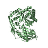
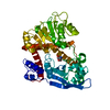
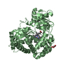
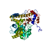
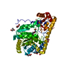
- PDB-6wms: Crystal Structure of Human REV-ERBbeta Ligand Binding Domain Co-B... -
+ Open data

- Basic information
| Entry | Database: PDB / ID: 6wms | ||||||
|---|---|---|---|---|---|---|---|
| Title | Crystal Structure of Human REV-ERBbeta Ligand Binding Domain Co-Bound to Heme and NCoR ID2 Peptide | ||||||
 Components Components |
| ||||||
 Keywords Keywords | TRANSCRIPTION / Nuclear receptor / heme-binding protein | ||||||
| Function / homology |  Function and homology information Function and homology informationregulation of skeletal muscle cell differentiation / Loss of MECP2 binding ability to the NCoR/SMRT complex / negative regulation of androgen receptor signaling pathway / circadian behavior / nuclear thyroid hormone receptor binding / negative regulation of glycolytic process / negative regulation of JNK cascade / NR1H2 & NR1H3 regulate gene expression to control bile acid homeostasis / negative regulation of fatty acid metabolic process / Notch-HLH transcription pathway ...regulation of skeletal muscle cell differentiation / Loss of MECP2 binding ability to the NCoR/SMRT complex / negative regulation of androgen receptor signaling pathway / circadian behavior / nuclear thyroid hormone receptor binding / negative regulation of glycolytic process / negative regulation of JNK cascade / NR1H2 & NR1H3 regulate gene expression to control bile acid homeostasis / negative regulation of fatty acid metabolic process / Notch-HLH transcription pathway / locomotor rhythm / histone deacetylase complex / lipid homeostasis / regulation of lipid metabolic process / Regulation of MECP2 expression and activity / Nuclear signaling by ERBB4 / NR1H3 & NR1H2 regulate gene expression linked to cholesterol transport and efflux / intracellular receptor signaling pathway / Regulation of lipid metabolism by PPARalpha / energy homeostasis / spindle assembly / transcription repressor complex / hormone-mediated signaling pathway / RORA,B,C and NR1D1 (REV-ERBA) regulate gene expression / Expression of BMAL (ARNTL), CLOCK, and NPAS2 / negative regulation of miRNA transcription / nuclear receptor binding / HDACs deacetylate histones / Heme signaling / PPARA activates gene expression / Transcriptional activation of mitochondrial biogenesis / Cytoprotection by HMOX1 / Downregulation of SMAD2/3:SMAD4 transcriptional activity / Nuclear Receptor transcription pathway / Transcriptional regulation of white adipocyte differentiation / regulation of circadian rhythm / DNA-binding transcription repressor activity, RNA polymerase II-specific / NOTCH1 Intracellular Domain Regulates Transcription / nuclear receptor activity / negative regulation of inflammatory response / Activation of anterior HOX genes in hindbrain development during early embryogenesis / Constitutive Signaling by NOTCH1 PEST Domain Mutants / Constitutive Signaling by NOTCH1 HD+PEST Domain Mutants / histone deacetylase binding / HCMV Early Events / sequence-specific double-stranded DNA binding / mitotic spindle / transcription corepressor activity / MLL4 and MLL3 complexes regulate expression of PPARG target genes in adipogenesis and hepatic steatosis / chromatin organization / regulation of inflammatory response / RNA polymerase II-specific DNA-binding transcription factor binding / DNA-binding transcription factor activity, RNA polymerase II-specific / cell differentiation / transcription cis-regulatory region binding / RNA polymerase II cis-regulatory region sequence-specific DNA binding / negative regulation of DNA-templated transcription / regulation of DNA-templated transcription / positive regulation of DNA-templated transcription / chromatin / negative regulation of transcription by RNA polymerase II / positive regulation of transcription by RNA polymerase II / zinc ion binding / nucleoplasm / membrane / nucleus / cytoplasm / cytosol Similarity search - Function | ||||||
| Biological species |  Homo sapiens (human) Homo sapiens (human) | ||||||
| Method |  X-RAY DIFFRACTION / SYNCHROTRON / MOLECULAR REPLACEMENT / Resolution: 2 Å X-RAY DIFFRACTION / SYNCHROTRON / MOLECULAR REPLACEMENT / Resolution: 2 Å | ||||||
 Authors Authors | Mosure, S.A. / Shang, J. / Kojetin, D.J. | ||||||
 Citation Citation | Journal: Sci Adv / Year: 2021 Title: Structural basis for heme-dependent NCoR binding to the transcriptional repressor REV-ERB beta. Authors: Mosure, S.A. / Strutzenberg, T.S. / Shang, J. / Munoz-Tello, P. / Solt, L.A. / Griffin, P.R. / Kojetin, D.J. | ||||||
| History |
|
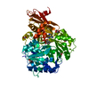
- Structure visualization
| Structure viewer | Molecule: MolmilJmol/JSmol |
|---|
- Downloads & links
-Download
| PDBx/mmCIF format | 6wms.cif.gz | 104.8 KB | Display | PDBx/mmCIF format |
|---|---|---|---|---|
| PDB format | pdb6wms.ent.gz | 78.2 KB | Display | PDB format |
| PDBx/mmJSON format | 6wms.json.gz | Tree view | PDBx/mmJSON format | |
| Others |  Other downloads Other downloads |
-Validation report
| Arichive directory | https://data.pdbj.org/pub/pdb/validation_reports/wm/6wmsftp://data.pdbj.org/pub/pdb/validation_reports/wm/6wms | HTTPS FTP |
|---|
-Related structure data
| Related structure data | 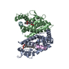 6wmqC 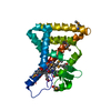 3cqvS S: Starting model for refinement C: citing same article ( |
|---|---|
| Similar structure data |


-Links
 PDBj
PDBj


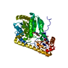
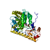
- Assembly
| Deposited unit | 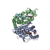
| ||||||||
|---|---|---|---|---|---|---|---|---|---|
| 1 |
| ||||||||
| Unit cell |
|
-Components
| #1: Protein | Mass: 22494.861 Da / Num. of mol.: 2 Source method: isolated from a genetically manipulated source Source: (gene. exp.) Homo sapiens (human) / Gene: NR1D2 / Production host:  #2: Protein/peptide | Mass: 2508.824 Da / Num. of mol.: 2 / Source method: obtained synthetically / Source: (synth.) Homo sapiens (human) / References: UniProt: Q86YY1, UniProt: O75376*PLUS#3: Chemical |   Mass: 616.487 Da / Num. of mol.: 2 / Source method: obtained synthetically / Formula: C34H32FeN4O4 / Feature type: SUBJECT OF INVESTIGATION Mass: 616.487 Da / Num. of mol.: 2 / Source method: obtained synthetically / Formula: C34H32FeN4O4 / Feature type: SUBJECT OF INVESTIGATION#4: Water | ChemComp-HOH / |  Mass: 18.015 Da / Num. of mol.: 204 / Source method: isolated from a natural source / Formula: H2O Mass: 18.015 Da / Num. of mol.: 204 / Source method: isolated from a natural source / Formula: H2OHas ligand of interest | Y | |
|---|
-Experimental details
-Experiment
| Experiment | Method: X-RAY DIFFRACTION / Number of used crystals: 1 |
|---|
- Sample preparation
| Crystal | Density Matthews: 2.38 Å3/Da / Density % sol: 48.36 % |
|---|---|
| Crystal grow | Temperature: 295 K / Method: vapor diffusion, sitting drop / Details: 0.2 M Mg formate dihydrate, 20% w/v PEG 3350 |
-Data collection
| Diffraction | Mean temperature: 100 K / Serial crystal experiment: N |
|---|---|
| Diffraction source | Source: SYNCHROTRON / Site: ALS  / Beamline: 5.0.2 / Wavelength: 0.97741 Å / Beamline: 5.0.2 / Wavelength: 0.97741 Å |
| Detector | Type: DECTRIS PILATUS3 S 6M / Detector: PIXEL / Date: Sep 9, 2019 |
| Radiation | Protocol: SINGLE WAVELENGTH / Monochromatic (M) / Laue (L): M / Scattering type: x-ray |
| Radiation wavelength | Wavelength: 0.97741 Å / Relative weight: 1 |
| Reflection | Resolution: 2→44 Å / Num. obs: 31527 / % possible obs: 99.09 % / Redundancy: 2 % / CC1/2: 1 / Net I/σ(I): 10.44 |
| Reflection shell | Resolution: 2→2.07 Å / Rmerge(I) obs: 0.313 / Mean I/σ(I) obs: 1.94 / Num. unique obs: 31527 / CC1/2: 0.754 / % possible all: 98.93 |
- Processing
| Software |
| ||||||||||||||||||||||||||||||||||||||||||||||||||||||||||||||||||||||||||||||||||||||||||
|---|---|---|---|---|---|---|---|---|---|---|---|---|---|---|---|---|---|---|---|---|---|---|---|---|---|---|---|---|---|---|---|---|---|---|---|---|---|---|---|---|---|---|---|---|---|---|---|---|---|---|---|---|---|---|---|---|---|---|---|---|---|---|---|---|---|---|---|---|---|---|---|---|---|---|---|---|---|---|---|---|---|---|---|---|---|---|---|---|---|---|---|
| Refinement | Method to determine structure: MOLECULAR REPLACEMENT Starting model: 3CQV Resolution: 2→43.699 Å / SU ML: 0.23 / Cross valid method: THROUGHOUT / σ(F): 1.36 / Phase error: 22.66
| ||||||||||||||||||||||||||||||||||||||||||||||||||||||||||||||||||||||||||||||||||||||||||
| Solvent computation | Shrinkage radii: 0.9 Å / VDW probe radii: 1.11 Å | ||||||||||||||||||||||||||||||||||||||||||||||||||||||||||||||||||||||||||||||||||||||||||
| Displacement parameters | Biso max: 91.43 Å2 / Biso mean: 32.6737 Å2 / Biso min: 12.42 Å2 | ||||||||||||||||||||||||||||||||||||||||||||||||||||||||||||||||||||||||||||||||||||||||||
| Refinement step | Cycle: final / Resolution: 2→43.699 Å
| ||||||||||||||||||||||||||||||||||||||||||||||||||||||||||||||||||||||||||||||||||||||||||
| Refine LS restraints |
| ||||||||||||||||||||||||||||||||||||||||||||||||||||||||||||||||||||||||||||||||||||||||||
| LS refinement shell | Refine-ID: X-RAY DIFFRACTION / Rfactor Rfree error: 0
|
