 Movie
Movie Controller

[English] 日本語
 Yorodumi
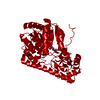
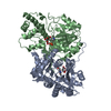
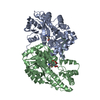
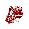
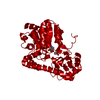
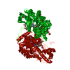
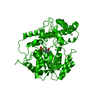
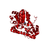
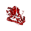
- PDB-1ase: THE STRUCTURE OF WILD TYPE E. COLI ASPARTATE AMINOTRANSFERASE REC... -
+ Open data

- Basic information
| Entry | Database: PDB / ID: 1ase | ||||||
|---|---|---|---|---|---|---|---|
| Title | THE STRUCTURE OF WILD TYPE E. COLI ASPARTATE AMINOTRANSFERASE RECONSTITUTED WITH PLP-N-OXIDE | ||||||
 Components Components | ASPARTATE AMINOTRANSFERASE | ||||||
 Keywords Keywords | AMINOTRANSFERASE | ||||||
| Function / homology |  Function and homology information Function and homology information: / L-tyrosine:2-oxoglutarate transaminase activity / L-phenylalanine biosynthetic process / aspartate transaminase / L-aspartate:2-oxoglutarate transaminase activity / pyridoxal phosphate binding / protein homodimerization activity / identical protein binding / cytoplasm / cytosol Similarity search - Function | ||||||
| Biological species |  | ||||||
| Method |  X-RAY DIFFRACTION / Resolution: 2.5 Å X-RAY DIFFRACTION / Resolution: 2.5 Å | ||||||
 Authors Authors | Schumacher, C. / Ringe, D. | ||||||
 Citation Citation | Journal: To be Published Title: The Structure of Wild Type E. Coli Aspartate Aminotransferase Reconstituted with Plp-N-Oxide Authors: Schumacher, C. / Ringe, D. #1: Journal: Biochemistry / Year: 1991Title: Activity and Structure of the Active Site Mutants R386Y and R386F of Escherichia Coli Aspartate Aminotransferase Authors: Danishefsky, A.T. / Onnufer, J.J. / Petsko, G.A. / Ringe, D. #2: Journal: Biochemistry / Year: 1989Title: 2.8 Angstroms Resolution Crystal Structure of an Active Site Mutant of Aspartate Aminotransferase from Escherichia Coli Authors: Smith, D.L. / Almo, S.C. / Toney, M.D. / Ringe, D. | ||||||
| History |
|
- Structure visualization
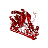
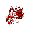
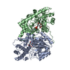
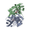
| Structure viewer | Molecule: MolmilJmol/JSmol |
|---|
- Downloads & links
-Download
| PDBx/mmCIF format | 1ase.cif.gz | 84.3 KB | Display | PDBx/mmCIF format |
|---|---|---|---|---|
| PDB format | pdb1ase.ent.gz | 63.1 KB | Display | PDB format |
| PDBx/mmJSON format | 1ase.json.gz | Tree view | PDBx/mmJSON format | |
| Others |  Other downloads Other downloads |
-Validation report
| Arichive directory | https://data.pdbj.org/pub/pdb/validation_reports/as/1aseftp://data.pdbj.org/pub/pdb/validation_reports/as/1ase | HTTPS FTP |
|---|
-Related structure data
| Similar structure data |
|---|
-Links
 PDBj
PDBj- Assembly
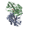
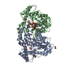
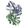
| Deposited unit | 
| ||||||||
|---|---|---|---|---|---|---|---|---|---|
| 1 | 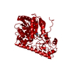
| ||||||||
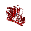
| Unit cell |
| ||||||||
| Atom site foot note | 1: CIS PROLINE - PRO 140 / 2: CIS PROLINE - PRO 196 | ||||||||
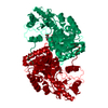
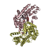
| Details | THE MOLECULE IS A DIMER. TO GENERATE THE OTHER CHAIN ONE MUST APPLY THE CRYSTALLOGRAPHIC SYMMETRY OPERATION (X, Y, 156.4-Z) TO THE COORDINATES IN THIS ENTRY. |
-Components
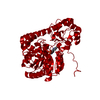
| #1: Protein | Mass: 43619.215 Da / Num. of mol.: 1 Source method: isolated from a genetically manipulated source Source: (gene. exp.) |
|---|---|
| #2: Chemical | ChemComp-NOP /   Mass: 263.141 Da / Num. of mol.: 1 / Source method: obtained synthetically / Formula: C8H10NO7P Mass: 263.141 Da / Num. of mol.: 1 / Source method: obtained synthetically / Formula: C8H10NO7P |
| #3: Chemical | ChemComp-MAE / 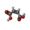  Mass: 116.072 Da / Num. of mol.: 1 / Source method: obtained synthetically / Formula: C4H4O4 Mass: 116.072 Da / Num. of mol.: 1 / Source method: obtained synthetically / Formula: C4H4O4 |
| #4: Water | ChemComp-HOH /  Mass: 18.015 Da / Num. of mol.: 23 / Source method: isolated from a natural source / Formula: H2O Mass: 18.015 Da / Num. of mol.: 23 / Source method: isolated from a natural source / Formula: H2O |
| Has protein modification | Y |
| Nonpolymer details | HET GROUP NOP 258 IS BOUND TO LYS 258 FORMING A PROTONATED SCHIFF BASE LINKAGE (BETWEEN NZ LYS 258 ...HET GROUP NOP 258 IS BOUND TO LYS 258 FORMING A PROTONATED |
-Experimental details
-Experiment
| Experiment | Method: X-RAY DIFFRACTION |
|---|
- Sample preparation
| Crystal | Density Matthews: 2.99 Å3/Da / Density % sol: 58.89 % |
|---|
-Data collection
| Radiation | Scattering type: x-ray |
|---|---|
| Radiation wavelength | Relative weight: 1 |
- Processing
| Software |
| ||||||||||||||||||||||||||||||||||||||||||||||||||||||||||||
|---|---|---|---|---|---|---|---|---|---|---|---|---|---|---|---|---|---|---|---|---|---|---|---|---|---|---|---|---|---|---|---|---|---|---|---|---|---|---|---|---|---|---|---|---|---|---|---|---|---|---|---|---|---|---|---|---|---|---|---|---|---|
| Refinement | Rfactor Rwork: 0.207 / Rfactor obs: 0.207 / Highest resolution: 2.5 Å | ||||||||||||||||||||||||||||||||||||||||||||||||||||||||||||
| Refinement step | Cycle: LAST / Highest resolution: 2.5 Å
| ||||||||||||||||||||||||||||||||||||||||||||||||||||||||||||
| Refine LS restraints |
|
