 Movie
Movie Controller

[English] 日本語
 Yorodumi
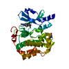
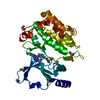
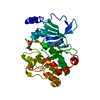
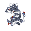
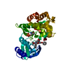
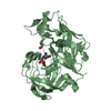
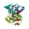
- PDB-6wly: PAK4 kinase domain in complex with LIMK1 Thr508 substrate peptide -
+ Open data

- Basic information
| Entry | Database: PDB / ID: 6wly | |||||||||
|---|---|---|---|---|---|---|---|---|---|---|
| Title | PAK4 kinase domain in complex with LIMK1 Thr508 substrate peptide | |||||||||
 Components Components |
| |||||||||
 Keywords Keywords | TRANSFERASE / serine/threonine kinase PAK4 / LIM kinase 1 / LIMK1 / phosphopeptide / Thr508 | |||||||||
| Function / homology |  Function and homology information Function and homology informationpositive regulation of actin filament bundle assembly / dendritic spine development / cadherin binding involved in cell-cell adhesion / positive regulation of focal adhesion disassembly / RHO GTPases Activate ROCKs / axon extension / Sema4D induced cell migration and growth-cone collapse / Activation of RAC1 / RHOV GTPase cycle / stress fiber assembly ...positive regulation of actin filament bundle assembly / dendritic spine development / cadherin binding involved in cell-cell adhesion / positive regulation of focal adhesion disassembly / RHO GTPases Activate ROCKs / axon extension / Sema4D induced cell migration and growth-cone collapse / Activation of RAC1 / RHOV GTPase cycle / stress fiber assembly / Fc-gamma receptor signaling pathway involved in phagocytosis / RHOJ GTPase cycle / RHOQ GTPase cycle / RHOU GTPase cycle / RHO GTPases activate PAKs / CDC42 GTPase cycle / regulation of MAPK cascade / Sema3A PAK dependent Axon repulsion / RHOG GTPase cycle / RHOH GTPase cycle / RAC3 GTPase cycle / RAC2 GTPase cycle / negative regulation of endothelial cell apoptotic process / ubiquitin ligase inhibitor activity / positive regulation of axon extension / Rho protein signal transduction / positive regulation of stress fiber assembly / heat shock protein binding / RAC1 GTPase cycle / EPHB-mediated forward signaling / cytoskeleton organization / cellular response to starvation / male germ cell nucleus / adherens junction / regulation of cell growth / Regulation of actin dynamics for phagocytic cup formation / positive regulation of angiogenesis / nervous system development / cell migration / lamellipodium / actin cytoskeleton organization / cytoskeleton / protein phosphorylation / protein-macromolecule adaptor activity / protein kinase activity / non-specific serine/threonine protein kinase / neuron projection / nuclear speck / intracellular signal transduction / protein stabilization / postsynapse / protein serine kinase activity / focal adhesion / protein serine/threonine kinase activity / apoptotic process / ubiquitin protein ligase binding / glutamatergic synapse / Golgi apparatus / signal transduction / ATP binding / membrane / metal ion binding / nucleus / cytoplasm / cytosol Similarity search - Function | |||||||||
| Biological species |  Homo sapiens (human) Homo sapiens (human) | |||||||||
| Method |  X-RAY DIFFRACTION / SYNCHROTRON / MOLECULAR REPLACEMENT / molecular replacement / Resolution: 1.9 Å X-RAY DIFFRACTION / SYNCHROTRON / MOLECULAR REPLACEMENT / molecular replacement / Resolution: 1.9 Å | |||||||||
 Authors Authors | Chetty, A.K. / Ha, B.H. / Boggon, T.J. | |||||||||
| Funding support |  United States, 2items United States, 2items
| |||||||||
 Citation Citation | Journal: J.Struct.Biol. / Year: 2020 Title: Recognition of physiological phosphorylation sites by p21-activated kinase 4. Authors: Chetty, A.K. / Sexton, J.A. / Ha, B.H. / Turk, B.E. / Boggon, T.J. | |||||||||
| History |
|
- Structure visualization
| Structure viewer | Molecule: MolmilJmol/JSmol |
|---|
- Downloads & links
-Download
| PDBx/mmCIF format | 6wly.cif.gz | 141 KB | Display | PDBx/mmCIF format |
|---|---|---|---|---|
| PDB format | pdb6wly.ent.gz | 107.8 KB | Display | PDB format |
| PDBx/mmJSON format | 6wly.json.gz | Tree view | PDBx/mmJSON format | |
| Others |  Other downloads Other downloads |
-Validation report
| Arichive directory | https://data.pdbj.org/pub/pdb/validation_reports/wl/6wlyftp://data.pdbj.org/pub/pdb/validation_reports/wl/6wly | HTTPS FTP |
|---|
-Related structure data
| Related structure data |  6wlxC  4fijS S: Starting model for refinement C: citing same article ( |
|---|---|
| Similar structure data | |
| Experimental dataset #1 | Data set type: diffraction image data / Metadata reference: 10.15785/SBGRID/782 |


-Links
 PDBj
PDBj


- Assembly
| Deposited unit | 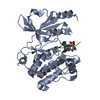
| ||||||||
|---|---|---|---|---|---|---|---|---|---|
| 1 |
| ||||||||
| Unit cell |
|
-Components
| #1: Protein | Mass: 39123.133 Da / Num. of mol.: 1 Source method: isolated from a genetically manipulated source Source: (gene. exp.) Homo sapiens (human) / Gene: PAK4, KIAA1142 / Production host:  References: UniProt: O96013, non-specific serine/threonine protein kinase |
|---|---|
| #2: Protein/peptide | Mass: 1224.456 Da / Num. of mol.: 1 / Source method: obtained synthetically / Source: (synth.) Homo sapiens (human)References: UniProt: P53667, non-specific serine/threonine protein kinase |
| #3: Water | ChemComp-HOH /  Mass: 18.015 Da / Num. of mol.: 135 / Source method: isolated from a natural source / Formula: H2O Mass: 18.015 Da / Num. of mol.: 135 / Source method: isolated from a natural source / Formula: H2O |
| Has ligand of interest | N |
| Has protein modification | Y |
-Experimental details
-Experiment
| Experiment | Method: X-RAY DIFFRACTION / Number of used crystals: 1 |
|---|
- Sample preparation
| Crystal | Density Matthews: 2.11 Å3/Da / Density % sol: 41.79 % |
|---|---|
| Crystal grow | Temperature: 295 K / Method: vapor diffusion, hanging drop / pH: 7.5 Details: 0.1M Tris-HCl, 2M Na acetate, pH 7.5, VAPOR DIFFUSION, HANGING DROP, temperature 295K, 2mM peptide |
-Data collection
| Diffraction | Mean temperature: 100 K / Serial crystal experiment: N | |||||||||||||||||||||||||||||||||||||||||||||||||||||||||||||||||||||||||||||||||||||||||||||||||||
|---|---|---|---|---|---|---|---|---|---|---|---|---|---|---|---|---|---|---|---|---|---|---|---|---|---|---|---|---|---|---|---|---|---|---|---|---|---|---|---|---|---|---|---|---|---|---|---|---|---|---|---|---|---|---|---|---|---|---|---|---|---|---|---|---|---|---|---|---|---|---|---|---|---|---|---|---|---|---|---|---|---|---|---|---|---|---|---|---|---|---|---|---|---|---|---|---|---|---|---|---|
| Diffraction source | Source: SYNCHROTRON / Site: APS / Beamline: 24-ID-E / Wavelength: 0.9792 Å | |||||||||||||||||||||||||||||||||||||||||||||||||||||||||||||||||||||||||||||||||||||||||||||||||||
| Detector | Type: DECTRIS EIGER X 16M / Detector: PIXEL / Date: Jul 7, 2018 | |||||||||||||||||||||||||||||||||||||||||||||||||||||||||||||||||||||||||||||||||||||||||||||||||||
| Radiation | Monochromator: Si(111) / Protocol: SINGLE WAVELENGTH / Monochromatic (M) / Laue (L): M / Scattering type: x-ray | |||||||||||||||||||||||||||||||||||||||||||||||||||||||||||||||||||||||||||||||||||||||||||||||||||
| Radiation wavelength | Wavelength: 0.9792 Å / Relative weight: 1 | |||||||||||||||||||||||||||||||||||||||||||||||||||||||||||||||||||||||||||||||||||||||||||||||||||
| Reflection | Resolution: 1.9→50 Å / Num. obs: 28404 / % possible obs: 99.9 % / Redundancy: 20.3 % / Rmerge(I) obs: 0.14 / Rpim(I) all: 0.03 / Rrim(I) all: 0.143 / Χ2: 0.984 / Net I/σ(I): 6.8 / Num. measured all: 576393 | |||||||||||||||||||||||||||||||||||||||||||||||||||||||||||||||||||||||||||||||||||||||||||||||||||
| Reflection shell | Diffraction-ID: 1
|
-Phasing
| Phasing | Method: molecular replacement | |||||||||
|---|---|---|---|---|---|---|---|---|---|---|
| Phasing MR |
|
- Processing
| Software |
| ||||||||||||||||||||||||||||||||||||||||||||||||||||||||||||||||||||||||||||||||||||||||||||||||||||
|---|---|---|---|---|---|---|---|---|---|---|---|---|---|---|---|---|---|---|---|---|---|---|---|---|---|---|---|---|---|---|---|---|---|---|---|---|---|---|---|---|---|---|---|---|---|---|---|---|---|---|---|---|---|---|---|---|---|---|---|---|---|---|---|---|---|---|---|---|---|---|---|---|---|---|---|---|---|---|---|---|---|---|---|---|---|---|---|---|---|---|---|---|---|---|---|---|---|---|---|---|---|
| Refinement | Method to determine structure: MOLECULAR REPLACEMENT Starting model: 4FIJ Resolution: 1.9→43.58 Å / SU ML: 0.2 / Cross valid method: THROUGHOUT / σ(F): 1.36 / Phase error: 22.39 / Stereochemistry target values: ML
| ||||||||||||||||||||||||||||||||||||||||||||||||||||||||||||||||||||||||||||||||||||||||||||||||||||
| Solvent computation | Shrinkage radii: 0.9 Å / VDW probe radii: 1.11 Å / Solvent model: FLAT BULK SOLVENT MODEL | ||||||||||||||||||||||||||||||||||||||||||||||||||||||||||||||||||||||||||||||||||||||||||||||||||||
| Displacement parameters | Biso max: 108.1 Å2 / Biso mean: 45.0861 Å2 / Biso min: 28.98 Å2 | ||||||||||||||||||||||||||||||||||||||||||||||||||||||||||||||||||||||||||||||||||||||||||||||||||||
| Refinement step | Cycle: final / Resolution: 1.9→43.58 Å
| ||||||||||||||||||||||||||||||||||||||||||||||||||||||||||||||||||||||||||||||||||||||||||||||||||||
| LS refinement shell | Refine-ID: X-RAY DIFFRACTION / Rfactor Rfree error: 0 / Total num. of bins used: 10 / % reflection obs: 100 %
| ||||||||||||||||||||||||||||||||||||||||||||||||||||||||||||||||||||||||||||||||||||||||||||||||||||
| Refinement TLS params. | Method: refined / Refine-ID: X-RAY DIFFRACTION
| ||||||||||||||||||||||||||||||||||||||||||||||||||||||||||||||||||||||||||||||||||||||||||||||||||||
| Refinement TLS group |
|
