 Movie
Movie Controller

[English] 日本語
 Yorodumi
- PDB-1ml4: The PALA-liganded Aspartate transcarbamoylase catalytic subunit f... -
+ Open data

- Basic information
| Entry | Database: PDB / ID: 1ml4 | ||||||
|---|---|---|---|---|---|---|---|
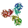
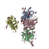
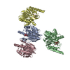
| Title | The PALA-liganded Aspartate transcarbamoylase catalytic subunit from Pyrococcus abyssi | ||||||
 Components Components | Aspartate Transcarbamoylase | ||||||
 Keywords Keywords | TRANSFERASE / BETA PLEATED SHEET / PROTEIN INHIBITOR COMPLEX | ||||||
| Function / homology |  Function and homology information Function and homology informationaspartate carbamoyltransferase / aspartate carbamoyltransferase activity / amino acid metabolic process / amino acid binding / 'de novo' UMP biosynthetic process / 'de novo' pyrimidine nucleobase biosynthetic process Similarity search - Function | ||||||
| Biological species |   Pyrococcus abyssi (archaea) Pyrococcus abyssi (archaea) | ||||||
| Method |  X-RAY DIFFRACTION / SYNCHROTRON / MOLECULAR REPLACEMENT / Resolution: 1.8 Å X-RAY DIFFRACTION / SYNCHROTRON / MOLECULAR REPLACEMENT / Resolution: 1.8 Å | ||||||
 Authors Authors | Van Boxstael, S. / Cunin, R. / Maes, D. | ||||||
 Citation Citation | Journal: J.Mol.Biol. / Year: 2003 Title: Aspartate Transcarbamylase from the Hyperthermophilic Archaeon Pyrococcus abyssi: Thermostability and 1.8A Resolution Crystal Structure of the Catalytic Subunit Complexed With the Bisubstrate ...Title: Aspartate Transcarbamylase from the Hyperthermophilic Archaeon Pyrococcus abyssi: Thermostability and 1.8A Resolution Crystal Structure of the Catalytic Subunit Complexed With the Bisubstrate Analogue N-Phosphonacetyl-L-aspartate. Authors: Van Boxstael, S. / Cunin, R. / Khan, S. / Maes, D. | ||||||
| History |
|
- Structure visualization
| Structure viewer | Molecule: MolmilJmol/JSmol |
|---|
- Downloads & links
-Download
| PDBx/mmCIF format | 1ml4.cif.gz | 75.4 KB | Display | PDBx/mmCIF format |
|---|---|---|---|---|
| PDB format | pdb1ml4.ent.gz | 55.8 KB | Display | PDB format |
| PDBx/mmJSON format | 1ml4.json.gz | Tree view | PDBx/mmJSON format | |
| Others |  Other downloads Other downloads |
-Validation report
| Arichive directory | https://data.pdbj.org/pub/pdb/validation_reports/ml/1ml4ftp://data.pdbj.org/pub/pdb/validation_reports/ml/1ml4 | HTTPS FTP |
|---|
-Related structure data
| Related structure data |  1ekxS S: Starting model for refinement |
|---|---|
| Similar structure data |


-Links
 PDBj
PDBj
- Assembly
| Deposited unit | 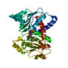
| |||||||||
|---|---|---|---|---|---|---|---|---|---|---|
| 1 | 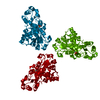
| |||||||||
| 2 |
| |||||||||
| 3 | 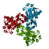
| |||||||||
| Unit cell |
| |||||||||
| Components on special symmetry positions |
| |||||||||
| Details | The biological assembly is a trimer generated by the operations : -Y,X-Y,Z and -X+Y,-X,Z |
-Components
| #1: Protein | Mass: 34954.379 Da / Num. of mol.: 1 Source method: isolated from a genetically manipulated source Source: (gene. exp.) Pyrococcus abyssi (archaea) / Gene: PyrB / Plasmid: Ptrc99A / Production host:  |
|---|---|
| #2: Chemical | ChemComp-PAL /   Mass: 255.119 Da / Num. of mol.: 1 / Source method: obtained synthetically / Formula: C6H10NO8P Mass: 255.119 Da / Num. of mol.: 1 / Source method: obtained synthetically / Formula: C6H10NO8P |
| #3: Water | ChemComp-HOH /  Mass: 18.015 Da / Num. of mol.: 77 / Source method: isolated from a natural source / Formula: H2O Mass: 18.015 Da / Num. of mol.: 77 / Source method: isolated from a natural source / Formula: H2O |
-Experimental details
-Experiment
| Experiment | Method: X-RAY DIFFRACTION / Number of used crystals: 1 |
|---|
- Sample preparation
| Crystal | Density Matthews: 2.52 Å3/Da / Density % sol: 50.87 % | ||||||||||||||||||||||||||||||||||||||||||
|---|---|---|---|---|---|---|---|---|---|---|---|---|---|---|---|---|---|---|---|---|---|---|---|---|---|---|---|---|---|---|---|---|---|---|---|---|---|---|---|---|---|---|---|
| Crystal grow | Temperature: 292 K / Method: vapor diffusion, hanging drop / pH: 6.8 Details: citric acid, pH 6.8, VAPOR DIFFUSION, HANGING DROP, temperature 292K | ||||||||||||||||||||||||||||||||||||||||||
| Crystal grow | *PLUS Temperature: 293 K / pH: 8.2 | ||||||||||||||||||||||||||||||||||||||||||
| Components of the solutions | *PLUS
|
-Data collection
| Diffraction | Mean temperature: 100 K |
|---|---|
| Diffraction source | Source: SYNCHROTRON / Site: EMBL/DESY, HAMBURG  / Beamline: BW7B / Wavelength: 0.84 / Beamline: BW7B / Wavelength: 0.84 |
| Detector | Type: MAR scanner 345 mm plate / Detector: IMAGE PLATE / Date: Sep 24, 2001 |
| Radiation | Monochromator: Triangular monochromator / Protocol: SINGLE WAVELENGTH / Monochromatic (M) / Laue (L): M / Scattering type: x-ray |
| Radiation wavelength | Wavelength: 0.84 Å / Relative weight: 1 |
| Reflection | Resolution: 1.8→25 Å / Num. all: 29667 / Num. obs: 29667 / % possible obs: 92.1 % / Observed criterion σ(F): 0 / Observed criterion σ(I): 0 / Redundancy: 2.38 % / Biso Wilson estimate: 20.9 Å2 / Rmerge(I) obs: 0.1 / Rsym value: 0.1 / Net I/σ(I): 10.69 |
| Reflection shell | Resolution: 1.8→1.86 Å / Redundancy: 1.9 % / Rmerge(I) obs: 0.33 / Mean I/σ(I) obs: 3.91 / Num. unique all: 2958 / Rsym value: 0.33 / % possible all: 93 |
| Reflection | *PLUS Lowest resolution: 25 Å / Num. measured all: 70669 / Rmerge(I) obs: 0.1 |
| Reflection shell | *PLUS Highest resolution: 1.8 Å / % possible obs: 93 % / Mean I/σ(I) obs: 3.9 |
- Processing
| Software |
| ||||||||||||||||||||||||||||||||||||
|---|---|---|---|---|---|---|---|---|---|---|---|---|---|---|---|---|---|---|---|---|---|---|---|---|---|---|---|---|---|---|---|---|---|---|---|---|---|
| Refinement | Method to determine structure: MOLECULAR REPLACEMENT Starting model: pdb 1EKX Resolution: 1.8→25 Å / Isotropic thermal model: ISOTROPIC / Cross valid method: Rfree / σ(F): 0 / σ(I): 0 / Stereochemistry target values: Engh & Huber / Details: CNS VERSION FOR HEMIHEDRAL TWINNED DATA
| ||||||||||||||||||||||||||||||||||||
| Displacement parameters | Biso mean: 27.45 Å2
| ||||||||||||||||||||||||||||||||||||
| Refinement step | Cycle: LAST / Resolution: 1.8→25 Å
| ||||||||||||||||||||||||||||||||||||
| Refine LS restraints |
| ||||||||||||||||||||||||||||||||||||
| LS refinement shell |
| ||||||||||||||||||||||||||||||||||||
| Xplor file |
| ||||||||||||||||||||||||||||||||||||
| Refinement | *PLUS Lowest resolution: 25 Å / Rfactor Rfree: 0.2118 / Rfactor Rwork: 0.1718 | ||||||||||||||||||||||||||||||||||||
| Solvent computation | *PLUS | ||||||||||||||||||||||||||||||||||||
| Displacement parameters | *PLUS | ||||||||||||||||||||||||||||||||||||
| Refine LS restraints | *PLUS
|
