 Movie
Movie Controller

[English] 日本語
 Yorodumi
- PDB-6wjl: Crystal structure of Glypican-2 core protein in complex with D3 Fab -
+ Open data

- Basic information
| Entry | Database: PDB / ID: 6wjl | |||||||||
|---|---|---|---|---|---|---|---|---|---|---|
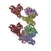
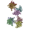
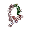
| Title | Crystal structure of Glypican-2 core protein in complex with D3 Fab | |||||||||
 Components Components |
| |||||||||
 Keywords Keywords | Oncoprotein/Immune System / Neuroblastoma / Oncoprotein / Proteoglycan / Therapeutic antibody / Oncoprotein-Immune System complex | |||||||||
| Function / homology |  Function and homology information Function and homology informationregulation of protein localization to membrane / Dengue Virus Attachment and Entry / Dengue Virus-Host Interactions / Defective B3GALT6 causes EDSP2 and SEMDJL1 / Defective B4GALT7 causes EDS, progeroid type / Defective B3GAT3 causes JDSSDHD / Defective EXT2 causes exostoses 2 / Defective EXT1 causes exostoses 1, TRPS2 and CHDS / Glycosaminoglycan-protein linkage region biosynthesis / HS-GAG biosynthesis ...regulation of protein localization to membrane / Dengue Virus Attachment and Entry / Dengue Virus-Host Interactions / Defective B3GALT6 causes EDSP2 and SEMDJL1 / Defective B4GALT7 causes EDS, progeroid type / Defective B3GAT3 causes JDSSDHD / Defective EXT2 causes exostoses 2 / Defective EXT1 causes exostoses 1, TRPS2 and CHDS / Glycosaminoglycan-protein linkage region biosynthesis / HS-GAG biosynthesis / HS-GAG degradation / smoothened signaling pathway / RSV-host interactions / Respiratory syncytial virus (RSV) attachment and entry / regulation of signal transduction / Retinoid metabolism and transport / side of membrane / lysosomal lumen / positive regulation of neuron projection development / Golgi lumen / neuron differentiation / cell migration / extracellular matrix / Attachment and Entry / synapse / cell surface / endoplasmic reticulum / extracellular region / plasma membrane Similarity search - Function | |||||||||
| Biological species |  Homo sapiens (human) Homo sapiens (human) | |||||||||
| Method |  X-RAY DIFFRACTION / SYNCHROTRON / MOLECULAR REPLACEMENT / Resolution: 3.3 Å X-RAY DIFFRACTION / SYNCHROTRON / MOLECULAR REPLACEMENT / Resolution: 3.3 Å | |||||||||
 Authors Authors | Raman, S. / Maris, J.M. / Bosse, K.R. / Julien, J.P. | |||||||||
| Funding support | 2items
| |||||||||
 Citation Citation | Journal: Cell Rep Med / Year: 2021 Title: A GPC2 antibody-drug conjugate is efficacious against neuroblastoma and small-cell lung cancer via binding a conformational epitope. Authors: Raman, S. / Buongervino, S.N. / Lane, M.V. / Zhelev, D.V. / Zhu, Z. / Cui, H. / Martinez, B. / Martinez, D. / Wang, Y. / Upton, K. / Patel, K. / Rathi, K.S. / Navia, C.T. / Harmon, D.B. / ...Authors: Raman, S. / Buongervino, S.N. / Lane, M.V. / Zhelev, D.V. / Zhu, Z. / Cui, H. / Martinez, B. / Martinez, D. / Wang, Y. / Upton, K. / Patel, K. / Rathi, K.S. / Navia, C.T. / Harmon, D.B. / Li, Y. / Pawel, B. / Dimitrov, D.S. / Maris, J.M. / Julien, J.P. / Bosse, K.R. | |||||||||
| History |
|
- Structure visualization
| Structure viewer | Molecule: MolmilJmol/JSmol |
|---|
- Downloads & links
-Download
| PDBx/mmCIF format | 6wjl.cif.gz | 369.1 KB | Display | PDBx/mmCIF format |
|---|---|---|---|---|
| PDB format | pdb6wjl.ent.gz | 292.8 KB | Display | PDB format |
| PDBx/mmJSON format | 6wjl.json.gz | Tree view | PDBx/mmJSON format | |
| Others |  Other downloads Other downloads |
-Validation report
| Arichive directory | https://data.pdbj.org/pub/pdb/validation_reports/wj/6wjlftp://data.pdbj.org/pub/pdb/validation_reports/wj/6wjl | HTTPS FTP |
|---|
-Related structure data
| Related structure data |  4acrS S: Starting model for refinement |
|---|---|
| Similar structure data |
-Links
 PDBj
PDBj


- Assembly
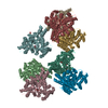
| Deposited unit | 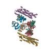
| ||||||||||
|---|---|---|---|---|---|---|---|---|---|---|---|
| 1 | 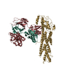
| ||||||||||
| 2 | 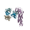
| ||||||||||
| Unit cell |
|
-Components
| #1: Protein | Mass: 53427.738 Da / Num. of mol.: 2 / Mutation: S55T, S92T, S155T Source method: isolated from a genetically manipulated source Source: (gene. exp.) Homo sapiens (human) / Gene: GPC2 / Production host: Homo sapiens (human) / References: UniProt: Q8N158#2: Antibody | Mass: 23499.227 Da / Num. of mol.: 2 Source method: isolated from a genetically manipulated source Source: (gene. exp.) Homo sapiens (human) / Production host: Homo sapiens (human)#3: Antibody | Mass: 11326.253 Da / Num. of mol.: 2 Source method: isolated from a genetically manipulated source Source: (gene. exp.) Production host: #4: Antibody | Mass: 23463.115 Da / Num. of mol.: 2 Source method: isolated from a genetically manipulated source Source: (gene. exp.) Homo sapiens (human) / Production host: Homo sapiens (human)Has protein modification | Y | |
|---|
-Experimental details
-Experiment
| Experiment | Method: X-RAY DIFFRACTION / Number of used crystals: 1 |
|---|
- Sample preparation
| Crystal | Density Matthews: 2.95 Å3/Da / Density % sol: 58.28 % |
|---|---|
| Crystal grow | Temperature: 293 K / Method: vapor diffusion, sitting drop Details: 0.2 M NaCl, 10% (w/v) PEG 3350 and 0.1 M disodium hydrogen phosphate |
-Data collection
| Diffraction | Mean temperature: 100 K / Serial crystal experiment: N |
|---|---|
| Diffraction source | Source: SYNCHROTRON / Site: APS  / Beamline: 23-ID-D / Wavelength: 1.03319 Å / Beamline: 23-ID-D / Wavelength: 1.03319 Å |
| Detector | Type: DECTRIS PILATUS3 6M / Detector: PIXEL / Date: Nov 13, 2018 |
| Radiation | Protocol: SINGLE WAVELENGTH / Monochromatic (M) / Laue (L): M / Scattering type: x-ray |
| Radiation wavelength | Wavelength: 1.03319 Å / Relative weight: 1 |
| Reflection | Resolution: 3.3→29.476 Å / Num. obs: 37835 / % possible obs: 99.76 % / Redundancy: 5.8 % / Biso Wilson estimate: 79.2 Å2 / CC1/2: 0.991 / Net I/σ(I): 7.3 |
| Reflection shell | Resolution: 3.3→3.418 Å / Num. unique obs: 3807 / CC1/2: 0.826 |
- Processing
| Software |
| ||||||||||||||||||||||||||||||||||||||||||||||||||||||||||||||||||||||
|---|---|---|---|---|---|---|---|---|---|---|---|---|---|---|---|---|---|---|---|---|---|---|---|---|---|---|---|---|---|---|---|---|---|---|---|---|---|---|---|---|---|---|---|---|---|---|---|---|---|---|---|---|---|---|---|---|---|---|---|---|---|---|---|---|---|---|---|---|---|---|---|
| Refinement | Method to determine structure: MOLECULAR REPLACEMENT Starting model: 4ACR Resolution: 3.3→29.476 Å / SU ML: 0.54 / Cross valid method: THROUGHOUT / σ(F): 1.37 / Phase error: 34.1 / Stereochemistry target values: ML
| ||||||||||||||||||||||||||||||||||||||||||||||||||||||||||||||||||||||
| Solvent computation | Shrinkage radii: 0.9 Å / VDW probe radii: 1.11 Å / Solvent model: FLAT BULK SOLVENT MODEL | ||||||||||||||||||||||||||||||||||||||||||||||||||||||||||||||||||||||
| Displacement parameters | Biso max: 159.22 Å2 / Biso mean: 72.9611 Å2 / Biso min: 23.99 Å2 | ||||||||||||||||||||||||||||||||||||||||||||||||||||||||||||||||||||||
| Refinement step | Cycle: final / Resolution: 3.3→29.476 Å
| ||||||||||||||||||||||||||||||||||||||||||||||||||||||||||||||||||||||
| LS refinement shell | Refine-ID: X-RAY DIFFRACTION / Rfactor Rfree error: 0 / % reflection obs: 100 %
|
