 Movie
Movie Controller


+ Open data

- Basic information
| Entry | Database: PDB / ID: 6wi6 | ||||||
|---|---|---|---|---|---|---|---|
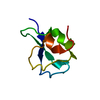
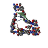
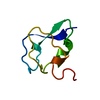
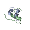
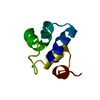
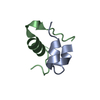
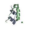
| Title | Crystal structure of plantacyclin B21AG | ||||||
 Components Components | Plantacyclin B21AG | ||||||
 Keywords Keywords | ANTIMICROBIAL PROTEIN / Circular bacteriocin / antimicrobial peptide / lactic acid bacteria | ||||||
| Function / homology | MALONATE ION Function and homology information Function and homology information | ||||||
| Biological species |  Lactobacillus plantarum (bacteria) Lactobacillus plantarum (bacteria) | ||||||
| Method |  X-RAY DIFFRACTION / SYNCHROTRON / AB INITIO PHASING / Resolution: 1.8 Å X-RAY DIFFRACTION / SYNCHROTRON / AB INITIO PHASING / Resolution: 1.8 Å | ||||||
 Authors Authors | Smith, A.T. / Gor, M.C. / Vezina, B. / McMahon, R. / King, G. / Panjikar, S. / Rehm, B. / Martin, J. | ||||||
| Funding support | 1items
| ||||||
 Citation Citation | Journal: Sci Rep / Year: 2020 Title: Crystal structure and site-directed mutagenesis of circular bacteriocin plantacyclin B21AG reveals cationic and aromatic residues important for antimicrobial activity. Authors: Gor, M.C. / Vezina, B. / McMahon, R.M. / King, G.J. / Panjikar, S. / Rehm, B.H.A. / Martin, J.L. / Smith, A.T. #1: Journal: Heliyon / Year: 2020 Title: Discovery and characterisation of circular bacteriocin plantacyclin B21AG from Lactiplantibacillus plantarum B21. Authors: Golneshin, A. / Gor, M.C. / Williamson, N. / Vezina, B. / Van, T.T.H. / May, B.K. / Smith, A.T. #2: Journal: Plos One / Year: 2020 Title: Cloning and functional expression of a food-grade circular bacteriocin, plantacyclin B21AG, in probiotic Lactobacillus plantarum WCFS1. Authors: Gor, M.C. / Golneshin, A. / Van, T.T.H. / Moore, R.J. / Smith, A.T. #3: Journal: Patent / Year: 2016Title: Bacteriocin polypeptides and uses thereof Authors: Golneshin, A. / Gor, M.C. / Van, T.T.H. / May, B.K. / Moore, R.J. / Smith, A.T. | ||||||
| History |
|
- Structure visualization
| Structure viewer | Molecule: MolmilJmol/JSmol |
|---|
- Downloads & links
-Download
| PDBx/mmCIF format | 6wi6.cif.gz | 33.7 KB | Display | PDBx/mmCIF format |
|---|---|---|---|---|
| PDB format | pdb6wi6.ent.gz | 22.1 KB | Display | PDB format |
| PDBx/mmJSON format | 6wi6.json.gz | Tree view | PDBx/mmJSON format | |
| Others |  Other downloads Other downloads |
-Validation report
| Arichive directory | https://data.pdbj.org/pub/pdb/validation_reports/wi/6wi6ftp://data.pdbj.org/pub/pdb/validation_reports/wi/6wi6 | HTTPS FTP |
|---|
-Related structure data
| Similar structure data |
|---|

-Links
 PDBj
PDBj- Assembly
| Deposited unit | 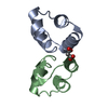
| ||||||||
|---|---|---|---|---|---|---|---|---|---|
| 1 | 
| ||||||||
| 2 | 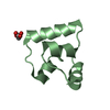
| ||||||||
| Unit cell |
| ||||||||
| Components on special symmetry positions |
|
-Components
| #1: Protein | Mass: 5689.649 Da / Num. of mol.: 2 / Source method: isolated from a natural source / Source: (natural) Lactobacillus plantarum (bacteria) / Strain: B21#2: Chemical | ChemComp-MLI / |   Mass: 102.046 Da / Num. of mol.: 1 / Source method: obtained synthetically / Formula: C3H2O4 Mass: 102.046 Da / Num. of mol.: 1 / Source method: obtained synthetically / Formula: C3H2O4#3: Water | ChemComp-HOH / |  Mass: 18.015 Da / Num. of mol.: 56 / Source method: isolated from a natural source / Formula: H2O Mass: 18.015 Da / Num. of mol.: 56 / Source method: isolated from a natural source / Formula: H2OHas ligand of interest | N | Has protein modification | Y | |
|---|
-Experimental details
-Experiment
| Experiment | Method: X-RAY DIFFRACTION / Number of used crystals: 1 |
|---|
- Sample preparation
| Crystal | Density Matthews: 2.24 Å3/Da / Density % sol: 44.98 % / Description: Cubic shape |
|---|---|
| Crystal grow | Temperature: 293.15 K / Method: vapor diffusion, hanging drop / pH: 7 Details: 1.1 M sodium malonate, 0.1 M HEPES pH7.0 and 0.5% v/v JED-2003 |
-Data collection
| Diffraction | Mean temperature: 100 K / Serial crystal experiment: N | ||||||||||||||||||||||||||||||
|---|---|---|---|---|---|---|---|---|---|---|---|---|---|---|---|---|---|---|---|---|---|---|---|---|---|---|---|---|---|---|---|
| Diffraction source | Source: SYNCHROTRON / Site: Australian Synchrotron  / Beamline: MX2 / Wavelength: 0.9537 Å / Beamline: MX2 / Wavelength: 0.9537 Å | ||||||||||||||||||||||||||||||
| Detector | Type: DECTRIS EIGER X 16M / Detector: PIXEL / Date: Oct 15, 2017 | ||||||||||||||||||||||||||||||
| Radiation | Protocol: SINGLE WAVELENGTH / Monochromatic (M) / Laue (L): M / Scattering type: x-ray | ||||||||||||||||||||||||||||||
| Radiation wavelength | Wavelength: 0.9537 Å / Relative weight: 1 | ||||||||||||||||||||||||||||||
| Reflection | Resolution: 1.8→46.58 Å / Num. obs: 9770 / % possible obs: 99.8 % / Redundancy: 13.1 % / CC1/2: 0.999 / Rmerge(I) obs: 0.076 / Rpim(I) all: 0.022 / Rrim(I) all: 0.079 / Net I/σ(I): 15.5 / Num. measured all: 128402 | ||||||||||||||||||||||||||||||
| Reflection shell | Diffraction-ID: 1
|
- Processing
| Software |
| ||||||||||||||||||||||||||||||||||||||||||||||||||||||||||||
|---|---|---|---|---|---|---|---|---|---|---|---|---|---|---|---|---|---|---|---|---|---|---|---|---|---|---|---|---|---|---|---|---|---|---|---|---|---|---|---|---|---|---|---|---|---|---|---|---|---|---|---|---|---|---|---|---|---|---|---|---|---|
| Refinement | Method to determine structure: AB INITIO PHASING / Resolution: 1.8→46.58 Å / Cor.coef. Fo:Fc: 0.973 / Cor.coef. Fo:Fc free: 0.955 / SU B: 3.187 / SU ML: 0.095 / Cross valid method: THROUGHOUT / σ(F): 0 / ESU R: 0.128 / ESU R Free: 0.127 / Stereochemistry target values: MAXIMUM LIKELIHOOD Details: HYDROGENS HAVE BEEN ADDED IN THE RIDING POSITIONS U VALUES : REFINED INDIVIDUALLY
| ||||||||||||||||||||||||||||||||||||||||||||||||||||||||||||
| Solvent computation | Ion probe radii: 0.8 Å / Shrinkage radii: 0.8 Å / VDW probe radii: 1.2 Å / Solvent model: MASK | ||||||||||||||||||||||||||||||||||||||||||||||||||||||||||||
| Displacement parameters | Biso max: 73.42 Å2 / Biso mean: 26.503 Å2 / Biso min: 15.88 Å2
| ||||||||||||||||||||||||||||||||||||||||||||||||||||||||||||
| Refinement step | Cycle: final / Resolution: 1.8→46.58 Å
| ||||||||||||||||||||||||||||||||||||||||||||||||||||||||||||
| Refine LS restraints |
| ||||||||||||||||||||||||||||||||||||||||||||||||||||||||||||
| LS refinement shell | Resolution: 1.803→1.849 Å / Rfactor Rfree error: 0 / Total num. of bins used: 20
|
