 ムービー
ムービー コントローラー


+ データを開く

- 基本情報
基本情報
| 登録情報 | データベース: PDB / ID: 6wbx | |||||||||||||||||||||
|---|---|---|---|---|---|---|---|---|---|---|---|---|---|---|---|---|---|---|---|---|---|---|
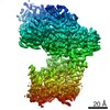
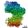
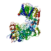
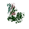
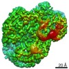
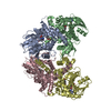
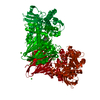
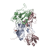
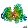
| タイトル | Single-Particle Cryo-EM Structure of Arabinofuranosyltransferase AftD from Mycobacteria, Mutant R1389S Class 1 | |||||||||||||||||||||
 要素 要素 | DUF3367 domain-containing protein | |||||||||||||||||||||
 キーワード キーワード | MEMBRANE PROTEIN / Glycosyltransferase / nanodisc / acyl carrier protein | |||||||||||||||||||||
| 機能・相同性 | Alpha-(1->3)-arabinofuranosyltransferase / Alpha-(1->3)-arabinofuranosyltransferase, N-terminal GT-C domain / Coagulation factors 5/8 type C domain (FA58C) profile. / Coagulation factor 5/8 C-terminal domain / Galactose-binding-like domain superfamily / transferase activity / membrane / DUF3367 domain-containing protein 機能・相同性情報 機能・相同性情報 | |||||||||||||||||||||
| 生物種 |  Mycobacteroides abscessus (バクテリア) Mycobacteroides abscessus (バクテリア) | |||||||||||||||||||||
| 手法 | 電子顕微鏡法 / 単粒子再構成法 / クライオ電子顕微鏡法 / 解像度: 3.5 Å | |||||||||||||||||||||
 データ登録者 データ登録者 | Tan, Y.Z. / Zhang, L. / Rodrigues, J. / Zheng, R.B. / Giacometti, S.I. / Rosario, A.L. / Kloss, B. / Dandey, V.P. / Wei, H. / Brunton, R. ...Tan, Y.Z. / Zhang, L. / Rodrigues, J. / Zheng, R.B. / Giacometti, S.I. / Rosario, A.L. / Kloss, B. / Dandey, V.P. / Wei, H. / Brunton, R. / Raczkowski, A.M. / Athayde, D. / Catalao, M.J. / Pimentel, M. / Clarke, O.B. / Lowary, T.L. / Archer, M. / Niederweis, M. / Potter, C.S. / Carragher, B. / Mancia, F. | |||||||||||||||||||||
| 資金援助 |  米国, 6件 米国, 6件
| |||||||||||||||||||||
 引用 引用 | ジャーナル: Mol Cell / 年: 2020 タイトル: Cryo-EM Structures and Regulation of Arabinofuranosyltransferase AftD from Mycobacteria. 著者: Yong Zi Tan / Lei Zhang / José Rodrigues / Ruixiang Blake Zheng / Sabrina I Giacometti / Ana L Rosário / Brian Kloss / Venkata P Dandey / Hui Wei / Richard Brunton / Ashleigh M Raczkowski / ...著者: Yong Zi Tan / Lei Zhang / José Rodrigues / Ruixiang Blake Zheng / Sabrina I Giacometti / Ana L Rosário / Brian Kloss / Venkata P Dandey / Hui Wei / Richard Brunton / Ashleigh M Raczkowski / Diogo Athayde / Maria João Catalão / Madalena Pimentel / Oliver B Clarke / Todd L Lowary / Margarida Archer / Michael Niederweis / Clinton S Potter / Bridget Carragher / Filippo Mancia /    要旨: Mycobacterium tuberculosis causes tuberculosis, a disease that kills over 1 million people each year. Its cell envelope is a common antibiotic target and has a unique structure due, in part, to two ...Mycobacterium tuberculosis causes tuberculosis, a disease that kills over 1 million people each year. Its cell envelope is a common antibiotic target and has a unique structure due, in part, to two lipidated polysaccharides-arabinogalactan and lipoarabinomannan. Arabinofuranosyltransferase D (AftD) is an essential enzyme involved in assembling these glycolipids. We present the 2.9-Å resolution structure of M. abscessus AftD, determined by single-particle cryo-electron microscopy. AftD has a conserved GT-C glycosyltransferase fold and three carbohydrate-binding modules. Glycan array analysis shows that AftD binds complex arabinose glycans. Additionally, AftD is non-covalently complexed with an acyl carrier protein (ACP). 3.4- and 3.5-Å structures of a mutant with impaired ACP binding reveal a conformational change, suggesting that ACP may regulate AftD function. Mutagenesis experiments using a conditional knockout constructed in M. smegmatis confirm the essentiality of the putative active site and the ACP binding for AftD function. | |||||||||||||||||||||
| 履歴 |
|
- 構造の表示
構造の表示
| ムービー |
ムービービューア |
|---|---|
| 構造ビューア | 分子: MolmilJmol/JSmol |
- ダウンロードとリンク
-ダウンロード
| PDBx/mmCIF形式 | 6wbx.cif.gz | 224.4 KB | 表示 | PDBx/mmCIF形式 |
|---|---|---|---|---|
| PDB形式 | pdb6wbx.ent.gz | 169.1 KB | 表示 | PDB形式 |
| PDBx/mmJSON形式 | 6wbx.json.gz | ツリー表示 | PDBx/mmJSON形式 | |
| その他 |  その他のダウンロード その他のダウンロード |
-検証レポート
| アーカイブディレクトリ | https://data.pdbj.org/pub/pdb/validation_reports/wb/6wbxftp://data.pdbj.org/pub/pdb/validation_reports/wb/6wbx | HTTPS FTP |
|---|
-関連構造データ
| 関連構造データ | 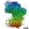 21600MC 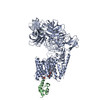 6w98C 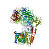 6wbyC C: 同じ文献を引用 ( M: このデータのモデリングに利用したマップデータ |
|---|---|
| 類似構造データ | |
| 電子顕微鏡画像生データ | EMPIAR-10391 (タイトル: Single-Particle Cryo-EM of Arabinofuranosyltransferase AftD from Mycobacteria, Mutant R1389S Data size: 2.3 TB Data #1: Unaligned and compressed multi-frame movies [micrographs - multiframe] Data #2: Aligned and dose-weighted micrographs [micrographs - single frame] Data #3: Final Particle Stacks with Refined Euler Angles and Shifts (Overall and Focused Refined on CBM3) [picked particles - single frame - processed]) |

-リンク
 PDBj
PDBj


- 集合体
集合体
| 登録構造単位 | 
|
|---|---|
| 1 |
|
-要素
| #1: タンパク質 | 分子量: 152818.641 Da / 分子数: 1 / 変異: R1389S / 由来タイプ: 組換発現 由来: (組換発現) Mycobacteroides abscessus (バクテリア)遺伝子: D2E76_26050 / 発現宿主: | ||||
|---|---|---|---|---|---|
| #2: 化合物 |   分子量: 40.078 Da / 分子数: 2 / 由来タイプ: 合成 / 式: Ca 分子量: 40.078 Da / 分子数: 2 / 由来タイプ: 合成 / 式: Ca研究の焦点であるリガンドがあるか | N | Has protein modification | Y | |
-実験情報
-実験
| 実験 | 手法: 電子顕微鏡法 |
|---|---|
| EM実験 | 試料の集合状態: PARTICLE / 3次元再構成法: 単粒子再構成法 |
- 試料調製
試料調製
| 構成要素 | 名称: Mutant R1389S Class 1 Mycobacterial Arabinofuranosyltransferase AftD Complexed with Acyl Carrier Protein タイプ: ORGANELLE OR CELLULAR COMPONENT / Entity ID: #1 / 由来: RECOMBINANT | ||||||||||||||||
|---|---|---|---|---|---|---|---|---|---|---|---|---|---|---|---|---|---|
| 分子量 |
| ||||||||||||||||
| 由来(天然) | 生物種: Mycobacteroides abscessus (バクテリア) | ||||||||||||||||
| 由来(組換発現) | 生物種: | ||||||||||||||||
| 緩衝液 | pH: 7.5 / 詳細: Solution was filtered and degassed. | ||||||||||||||||
| 緩衝液成分 |
| ||||||||||||||||
| 試料 | 濃度: 1 mg/ml / 包埋: NO / シャドウイング: NO / 染色: NO / 凍結: YES / 詳細: Protein was incorporated into lipid nanodiscs. | ||||||||||||||||
| 試料支持 | グリッドの材料: GOLD / グリッドのタイプ: UltrAuFoil | ||||||||||||||||
| 急速凍結 | 装置: LEICA EM GP / 凍結剤: ETHANE / 凍結前の試料温度: 298 K |
- 電子顕微鏡撮影
| 実験機器 |  モデル: Titan Krios / 画像提供: FEI Company |
|---|---|
| 顕微鏡 | モデル: FEI TITAN KRIOS |
| 電子銃 | 電子線源:  FIELD EMISSION GUN / 加速電圧: 300 kV / 照射モード: FLOOD BEAM FIELD EMISSION GUN / 加速電圧: 300 kV / 照射モード: FLOOD BEAM |
| 電子レンズ | モード: BRIGHT FIELD / 倍率(公称値): 130000 X / 最大 デフォーカス(公称値): 2000 nm / 最小 デフォーカス(公称値): 1000 nm / Cs: 2.7 mm / アライメント法: ZEMLIN TABLEAU |
| 試料ホルダ | 凍結剤: NITROGEN 試料ホルダーモデル: FEI TITAN KRIOS AUTOGRID HOLDER |
| 撮影 | 平均露光時間: 13.05 sec. / 電子線照射量: 96.78 e/Å2 / 検出モード: COUNTING フィルム・検出器のモデル: GATAN K2 QUANTUM (4k x 4k) 撮影したグリッド数: 1 / 実像数: 4886 |
| 電子光学装置 | エネルギーフィルター名称: GIF Bioquantum / エネルギーフィルタースリット幅: 15 eV |
| 画像スキャン | サンプリングサイズ: 5 µm / 横: 3838 / 縦: 3710 / 動画フレーム数/画像: 90 / 利用したフレーム数/画像: 1-90 |
- 解析
解析
| EMソフトウェア |
| ||||||||||||||||||||||||||||||||||||||||||||
|---|---|---|---|---|---|---|---|---|---|---|---|---|---|---|---|---|---|---|---|---|---|---|---|---|---|---|---|---|---|---|---|---|---|---|---|---|---|---|---|---|---|---|---|---|---|
| 画像処理 | 詳細: Energy filter slit width of 15 eV was used during the collection and was aligned automatically every hour using Leginon. | ||||||||||||||||||||||||||||||||||||||||||||
| CTF補正 | 詳細: CTF was estimated using GCTF and refined throughout the pipeline using cisTEM. タイプ: PHASE FLIPPING AND AMPLITUDE CORRECTION | ||||||||||||||||||||||||||||||||||||||||||||
| 粒子像の選択 | 選択した粒子像数: 226478 | ||||||||||||||||||||||||||||||||||||||||||||
| 対称性 | 点対称性: C1 (非対称) | ||||||||||||||||||||||||||||||||||||||||||||
| 3次元再構成 | 解像度: 3.5 Å / 解像度の算出法: FSC 0.143 CUT-OFF / 粒子像の数: 37814 / クラス平均像の数: 1 / 対称性のタイプ: POINT | ||||||||||||||||||||||||||||||||||||||||||||
| 原子モデル構築 | プロトコル: AB INITIO MODEL / 空間: REAL | ||||||||||||||||||||||||||||||||||||||||||||
| 原子モデル構築 | PDB-ID: 6W98 PDB chain-ID: A / Accession code: 6W98 / Source name: PDB / タイプ: experimental model |
