 Movie
Movie Controller

[English] 日本語
 Yorodumi
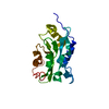
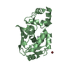
- PDB-6w9o: Crystal structure of an OTU deubiquitinase from Wolbachia pipient... -
+ Open data

- Basic information
| Entry | Database: PDB / ID: 6w9o | |||||||||
|---|---|---|---|---|---|---|---|---|---|---|
| Title | Crystal structure of an OTU deubiquitinase from Wolbachia pipientis wMel | |||||||||
 Components Components | OTU domain-containing protein wMelOTU | |||||||||
 Keywords Keywords | HYDROLASE / Deubiquitinase / Bacterial effector | |||||||||
| Function / homology | OTU domain / OTU domain profile. / ACETATE ION / OTU domain-containing protein Function and homology information Function and homology information | |||||||||
| Biological species |  Wolbachia pipientis wMel (bacteria) Wolbachia pipientis wMel (bacteria) | |||||||||
| Method |  X-RAY DIFFRACTION / SYNCHROTRON / MOLECULAR REPLACEMENT / molecular replacement / Resolution: 1.47 Å X-RAY DIFFRACTION / SYNCHROTRON / MOLECULAR REPLACEMENT / molecular replacement / Resolution: 1.47 Å | |||||||||
 Authors Authors | Schubert, A.F. / Pruneda, J.N. / Komander, D. | |||||||||
| Funding support |  United Kingdom, European Union, 2items United Kingdom, European Union, 2items
| |||||||||
 Citation Citation | Journal: Embo J. / Year: 2020 Title: Identification and characterization of diverse OTU deubiquitinases in bacteria. Authors: Schubert, A.F. / Nguyen, J.V. / Franklin, T.G. / Geurink, P.P. / Roberts, C.G. / Sanderson, D.J. / Miller, L.N. / Ovaa, H. / Hofmann, K. / Pruneda, J.N. / Komander, D. | |||||||||
| History |
|
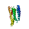
- Structure visualization
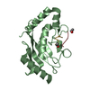
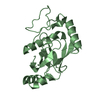
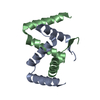
| Structure viewer | Molecule: MolmilJmol/JSmol |
|---|
- Downloads & links
-Download
| PDBx/mmCIF format | 6w9o.cif.gz | 90.1 KB | Display | PDBx/mmCIF format |
|---|---|---|---|---|
| PDB format | pdb6w9o.ent.gz | 65.9 KB | Display | PDB format |
| PDBx/mmJSON format | 6w9o.json.gz | Tree view | PDBx/mmJSON format | |
| Others |  Other downloads Other downloads |
-Validation report
| Summary document | 6w9o_validation.pdf.gz | 441.9 KB | Display | wwPDB validaton report |
|---|---|---|---|---|
| Full document | 6w9o_full_validation.pdf.gz | 443 KB | Display | |
| Data in XML | 6w9o_validation.xml.gz | 10.5 KB | Display | |
| Data in CIF | 6w9o_validation.cif.gz | 14.7 KB | Display | |
| Arichive directory | https://data.pdbj.org/pub/pdb/validation_reports/w9/6w9oftp://data.pdbj.org/pub/pdb/validation_reports/w9/6w9o | HTTPS FTP |
-Related structure data
| Related structure data |  6w9rC  6w9sC  3c0rS S: Starting model for refinement C: citing same article ( |
|---|---|
| Similar structure data |


-Links
 PDBj
PDBj- Assembly
| Deposited unit | 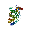
| ||||||||
|---|---|---|---|---|---|---|---|---|---|
| 1 |
| ||||||||
| Unit cell |
|
-Components
| #1: Protein | Mass: 24488.027 Da / Num. of mol.: 1 Source method: isolated from a genetically manipulated source Source: (gene. exp.) Wolbachia pipientis wMel (bacteria) / Gene: WD_0443 / Production host: |
|---|---|
| #2: Chemical | ChemComp-ACT /   Mass: 59.044 Da / Num. of mol.: 1 / Source method: obtained synthetically / Formula: C2H3O2 Mass: 59.044 Da / Num. of mol.: 1 / Source method: obtained synthetically / Formula: C2H3O2 |
| #3: Water | ChemComp-HOH /  Mass: 18.015 Da / Num. of mol.: 184 / Source method: isolated from a natural source / Formula: H2O Mass: 18.015 Da / Num. of mol.: 184 / Source method: isolated from a natural source / Formula: H2O |
| Has ligand of interest | N |
-Experimental details
-Experiment
| Experiment | Method: X-RAY DIFFRACTION / Number of used crystals: 1 |
|---|
- Sample preparation
| Crystal | Density Matthews: 1.94 Å3/Da / Density % sol: 36.7 % |
|---|---|
| Crystal grow | Temperature: 291 K / Method: vapor diffusion, sitting drop Details: 0.2 M sodium acetate, 32% PEG 4K, 0.1 M Tris pH 8.5 |
-Data collection
| Diffraction | Mean temperature: 100 K / Serial crystal experiment: N | ||||||||||||||||||||||||||||||
|---|---|---|---|---|---|---|---|---|---|---|---|---|---|---|---|---|---|---|---|---|---|---|---|---|---|---|---|---|---|---|---|
| Diffraction source | Source: SYNCHROTRON / Site: Diamond / Beamline: I24 / Wavelength: 0.96862 Å | ||||||||||||||||||||||||||||||
| Detector | Type: DECTRIS PILATUS 6M / Detector: PIXEL / Date: Jun 28, 2014 | ||||||||||||||||||||||||||||||
| Radiation | Protocol: SINGLE WAVELENGTH / Monochromatic (M) / Laue (L): M / Scattering type: x-ray | ||||||||||||||||||||||||||||||
| Radiation wavelength | Wavelength: 0.96862 Å / Relative weight: 1 | ||||||||||||||||||||||||||||||
| Reflection | Resolution: 1.47→33 Å / Num. obs: 33057 / % possible obs: 99.7 % / Redundancy: 4.3 % / CC1/2: 0.999 / Rmerge(I) obs: 0.053 / Rpim(I) all: 0.028 / Rrim(I) all: 0.061 / Net I/σ(I): 15.4 / Num. measured all: 141445 / Scaling rejects: 13 | ||||||||||||||||||||||||||||||
| Reflection shell | Diffraction-ID: 1
|
-Phasing
| Phasing | Method: molecular replacement |
|---|
- Processing
| Software |
| |||||||||||||||||||||||||||||||||||||||||||||||||||||||||||||||||||||||||||||||||||||||||||
|---|---|---|---|---|---|---|---|---|---|---|---|---|---|---|---|---|---|---|---|---|---|---|---|---|---|---|---|---|---|---|---|---|---|---|---|---|---|---|---|---|---|---|---|---|---|---|---|---|---|---|---|---|---|---|---|---|---|---|---|---|---|---|---|---|---|---|---|---|---|---|---|---|---|---|---|---|---|---|---|---|---|---|---|---|---|---|---|---|---|---|---|---|
| Refinement | Method to determine structure: MOLECULAR REPLACEMENT Starting model: 3C0R Resolution: 1.47→33 Å / SU ML: 0.13 / Cross valid method: THROUGHOUT / σ(F): 1.33 / Phase error: 19.48
| |||||||||||||||||||||||||||||||||||||||||||||||||||||||||||||||||||||||||||||||||||||||||||
| Solvent computation | Shrinkage radii: 0.9 Å / VDW probe radii: 1.11 Å | |||||||||||||||||||||||||||||||||||||||||||||||||||||||||||||||||||||||||||||||||||||||||||
| Displacement parameters | Biso max: 110.48 Å2 / Biso mean: 27.0821 Å2 / Biso min: 10.45 Å2 | |||||||||||||||||||||||||||||||||||||||||||||||||||||||||||||||||||||||||||||||||||||||||||
| Refinement step | Cycle: final / Resolution: 1.47→33 Å
| |||||||||||||||||||||||||||||||||||||||||||||||||||||||||||||||||||||||||||||||||||||||||||
| LS refinement shell | Refine-ID: X-RAY DIFFRACTION / Rfactor Rfree error: 0 / Total num. of bins used: 12
| |||||||||||||||||||||||||||||||||||||||||||||||||||||||||||||||||||||||||||||||||||||||||||
| Refinement TLS params. | Method: refined / Origin x: -5.6051 Å / Origin y: -11.0613 Å / Origin z: 19.9715 Å
| |||||||||||||||||||||||||||||||||||||||||||||||||||||||||||||||||||||||||||||||||||||||||||
| Refinement TLS group |
|
