Evidence: assay for oligomerization, Actin polymerizes in the presence of polymerizing buffer. This actin polymerization is severely inhibited in the presence of the ligand (synthetic toxin).
Type
Name
Symmetry operation
Number
identity operation
1_555
x,y,z
1
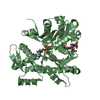
Buried area
2540 Å2
ΔGint
-12 kcal/mol
Surface area
15200 Å2
Method
PISA
Unit cell
Length a, b, c (Å)
37.763, 74.869, 61.197
Angle α, β, γ (deg.)
90.000, 93.023, 90.000
Int Tables number
4
Space group name H-M
P1211
Space group name Hall
P2yb
-
Components
-
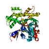
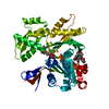
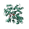
Protein , 1 types, 1 molecules A
#1: Protein
Actin, alphaskeletalmuscle / Alpha-actin-1
Mass: 42096.953 Da / Num. of mol.: 1 / Source method: isolated from a natural source / Source: (natural) Oryctolagus cuniculus (rabbit) / Tissue: heart / References: UniProt: P68135
In the structure databanks used in Yorodumi, some data are registered as the other names, "COVID-19 virus" and "2019-nCoV". Here are the details of the virus and the list of structure data.
Jan 31, 2019. EMDB accession codes are about to change! (news from PDBe EMDB page)
EMDB accession codes are about to change! (news from PDBe EMDB page)
The allocation of 4 digits for EMDB accession codes will soon come to an end. Whilst these codes will remain in use, new EMDB accession codes will include an additional digit and will expand incrementally as the available range of codes is exhausted. The current 4-digit format prefixed with “EMD-” (i.e. EMD-XXXX) will advance to a 5-digit format (i.e. EMD-XXXXX), and so on. It is currently estimated that the 4-digit codes will be depleted around Spring 2019, at which point the 5-digit format will come into force.
The EM Navigator/Yorodumi systems omit the EMD- prefix.
Related info.:Q: What is EMD? / ID/Accession-code notation in Yorodumi/EM Navigator
Yorodumi is a browser for structure data from EMDB, PDB, SASBDB, etc.
This page is also the successor to EM Navigator detail page, and also detail information page/front-end page for Omokage search.
The word "yorodu" (or yorozu) is an old Japanese word meaning "ten thousand". "mi" (miru) is to see.
Related info.:EMDB / PDB / SASBDB / Comparison of 3 databanks / Yorodumi Search / Aug 31, 2016. New EM Navigator & Yorodumi / Yorodumi Papers / Jmol/JSmol / Function and homology information / Changes in new EM Navigator and Yorodumi
 Movie
Movie Controller

 Yorodumi
 Open data
 Basic information
 Components
 Keywords
 Function and homology information
 X-RAY DIFFRACTION /
  Authors
 Canada, 1items
  Citation
 Structure visualization
 Downloads & links
 Other downloads

 PDBj
PDBj


 Assembly


 Mass: 40.078 Da / Num. of mol.: 1 / Source method: obtained synthetically / Formula: Ca
 Mass: 507.181 Da / Num. of mol.: 1 / Source method: obtained synthetically / Formula: C10H16N5O13P3 / Comment: ATP, energy-carrying molecule*YM
 Mass: 395.513 Da / Num. of mol.: 1 / Source method: obtained synthetically / Formula: C20H29NO5S / Comment: toxin*YM
 Mass: 62.068 Da / Num. of mol.: 3 / Source method: obtained synthetically / Formula: C2H6O2
 Mass: 598.812 Da / Num. of mol.: 1 / Source method: obtained synthetically / Formula: C32H58N2O8 / Feature type: SUBJECT OF INVESTIGATION
 Sample preparation
 Processing