 Movie
Movie Controller


+ Open data

- Basic information
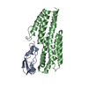
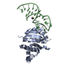
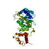
| Entry | Database: PDB / ID: 6w67 | ||||||
|---|---|---|---|---|---|---|---|
| Title | The structure of S172A Keap1-BTB domain | ||||||
 Components Components | Kelch-like ECH-associated protein 1 | ||||||
 Keywords Keywords | SIGNALING PROTEIN / BTB domain | ||||||
| Function / homology |  Function and homology information Function and homology informationNuclear events mediated by NFE2L2 / negative regulation of response to oxidative stress / Cul3-RING ubiquitin ligase complex / transcription regulator inhibitor activity / ubiquitin-like ligase-substrate adaptor activity / inclusion body / regulation of autophagy / actin filament / disordered domain specific binding / KEAP1-NFE2L2 pathway ...Nuclear events mediated by NFE2L2 / negative regulation of response to oxidative stress / Cul3-RING ubiquitin ligase complex / transcription regulator inhibitor activity / ubiquitin-like ligase-substrate adaptor activity / inclusion body / regulation of autophagy / actin filament / disordered domain specific binding / KEAP1-NFE2L2 pathway / positive regulation of proteasomal ubiquitin-dependent protein catabolic process / Antigen processing: Ubiquitination & Proteasome degradation / Neddylation / cellular response to oxidative stress / midbody / Potential therapeutics for SARS / ubiquitin-dependent protein catabolic process / RNA polymerase II-specific DNA-binding transcription factor binding / proteasome-mediated ubiquitin-dependent protein catabolic process / Ub-specific processing proteases / protein ubiquitination / negative regulation of transcription by RNA polymerase II / endoplasmic reticulum / nucleoplasm / identical protein binding / cytosol / cytoplasm Similarity search - Function | ||||||
| Biological species |  Homo sapiens (human) Homo sapiens (human) | ||||||
| Method |  X-RAY DIFFRACTION / SYNCHROTRON / FOURIER SYNTHESIS / Resolution: 2.2 Å X-RAY DIFFRACTION / SYNCHROTRON / FOURIER SYNTHESIS / Resolution: 2.2 Å | ||||||
 Authors Authors | Mena, E.L. / Gee, C.L. / Kuriyan, J. / Rape, M. | ||||||
| Funding support |  United States, 1items United States, 1items
| ||||||
 Citation Citation | Journal: Nature / Year: 2020 Title: Structural basis for dimerization quality control. Authors: Elijah L Mena / Predrag Jevtić / Basil J Greber / Christine L Gee / Brandon G Lew / David Akopian / Eva Nogales / John Kuriyan / Michael Rape / Abstract: Most quality control pathways target misfolded proteins to prevent toxic aggregation and neurodegeneration. Dimerization quality control further improves proteostasis by eliminating complexes of ...Most quality control pathways target misfolded proteins to prevent toxic aggregation and neurodegeneration. Dimerization quality control further improves proteostasis by eliminating complexes of aberrant composition, but how it detects incorrect subunits remains unknown. Here we provide structural insight into target selection by SCF-FBXL17, a dimerization-quality-control E3 ligase that ubiquitylates and helps to degrade inactive heterodimers of BTB proteins while sparing functional homodimers. We find that SCF-FBXL17 disrupts aberrant BTB dimers that fail to stabilize an intermolecular β-sheet around a highly divergent β-strand of the BTB domain. Complex dissociation allows SCF-FBXL17 to wrap around a single BTB domain, resulting in robust ubiquitylation. SCF-FBXL17 therefore probes both shape and complementarity of BTB domains, a mechanism that is well suited to establish quality control of complex composition for recurrent interaction modules. | ||||||
| History |
|
- Structure visualization
| Structure viewer | Molecule: MolmilJmol/JSmol |
|---|
- Downloads & links
-Download
| PDBx/mmCIF format | 6w67.cif.gz | 40 KB | Display | PDBx/mmCIF format |
|---|---|---|---|---|
| PDB format | pdb6w67.ent.gz | 25.5 KB | Display | PDB format |
| PDBx/mmJSON format | 6w67.json.gz | Tree view | PDBx/mmJSON format | |
| Others |  Other downloads Other downloads |
-Validation report
| Arichive directory | https://data.pdbj.org/pub/pdb/validation_reports/w6/6w67ftp://data.pdbj.org/pub/pdb/validation_reports/w6/6w67 | HTTPS FTP |
|---|
-Related structure data
| Related structure data | 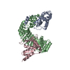 6w66C  6w68C 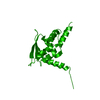 6w69C 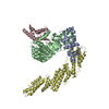 6wcqC 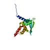 4cxiS S: Starting model for refinement C: citing same article ( |
|---|---|
| Similar structure data |


-Links
 PDBj
PDBj


- Assembly
| Deposited unit | 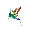
| ||||||||
|---|---|---|---|---|---|---|---|---|---|
| 1 | 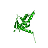
| ||||||||
| Unit cell |
|
-Components
| #1: Protein | Mass: 14937.227 Da / Num. of mol.: 1 / Fragment: BTB domain / Mutation: S172A Source method: isolated from a genetically manipulated source Source: (gene. exp.) Homo sapiens (human) / Gene: KEAP1, INRF2, KIAA0132, KLHL19 / Production host:  |
|---|---|
| #2: Water | ChemComp-HOH /  Mass: 18.015 Da / Num. of mol.: 23 / Source method: isolated from a natural source / Formula: H2O Mass: 18.015 Da / Num. of mol.: 23 / Source method: isolated from a natural source / Formula: H2O |
-Experimental details
-Experiment
| Experiment | Method: X-RAY DIFFRACTION / Number of used crystals: 1 |
|---|
- Sample preparation
| Crystal | Density Matthews: 2.34 Å3/Da / Density % sol: 47.51 % |
|---|---|
| Crystal grow | Temperature: 293 K / Method: vapor diffusion, hanging drop Details: 160-400 mM lithium acetate and 14-18% PEG 3350 1:1 with protein at 11mg/ml in 150 mM NaCl, 25 mM Tris-HCl pH 8.0, 1 mM TCEP |
-Data collection
| Diffraction | Mean temperature: 100 K / Serial crystal experiment: N | ||||||||||||||||||||||||||||||
|---|---|---|---|---|---|---|---|---|---|---|---|---|---|---|---|---|---|---|---|---|---|---|---|---|---|---|---|---|---|---|---|
| Diffraction source | Source: SYNCHROTRON / Site: ALS / Beamline: 8.3.1 / Wavelength: 1.11583 Å | ||||||||||||||||||||||||||||||
| Detector | Type: DECTRIS PILATUS 6M / Detector: PIXEL / Date: Aug 23, 2018 | ||||||||||||||||||||||||||||||
| Radiation | Monochromator: S111 / Protocol: SINGLE WAVELENGTH / Monochromatic (M) / Laue (L): M / Scattering type: x-ray | ||||||||||||||||||||||||||||||
| Radiation wavelength | Wavelength: 1.11583 Å / Relative weight: 1 | ||||||||||||||||||||||||||||||
| Reflection | Resolution: 2.2→44.373 Å / Num. obs: 8245 / % possible obs: 100 % / Redundancy: 24.5 % / CC1/2: 1 / Rmerge(I) obs: 0.198 / Rpim(I) all: 0.04 / Rrim(I) all: 0.202 / Net I/σ(I): 16.1 | ||||||||||||||||||||||||||||||
| Reflection shell | Diffraction-ID: 1
|
- Processing
| Software |
| ||||||||||||||||||||||||
|---|---|---|---|---|---|---|---|---|---|---|---|---|---|---|---|---|---|---|---|---|---|---|---|---|---|
| Refinement | Method to determine structure: FOURIER SYNTHESIS Starting model: 4CXI Resolution: 2.2→44.37 Å / SU ML: 0.3 / Cross valid method: THROUGHOUT / σ(F): 1.34 / Phase error: 35.11
| ||||||||||||||||||||||||
| Solvent computation | Shrinkage radii: 0.9 Å / VDW probe radii: 1.11 Å | ||||||||||||||||||||||||
| Displacement parameters | Biso max: 111.29 Å2 / Biso mean: 55.819 Å2 / Biso min: 29.27 Å2 | ||||||||||||||||||||||||
| Refinement step | Cycle: final / Resolution: 2.2→44.37 Å
| ||||||||||||||||||||||||
| Refine LS restraints |
| ||||||||||||||||||||||||
| LS refinement shell | Refine-ID: X-RAY DIFFRACTION / Rfactor Rfree error: 0 / % reflection obs: 100 %
|
