 Movie
Movie Controller

[English] 日本語
 Yorodumi
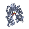
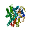
- PDB-6w0s: Crystal structure of substrate free cytochrome P450 NasF5053 from... -
+ Open data

- Basic information
| Entry | Database: PDB / ID: 6w0s | |||||||||
|---|---|---|---|---|---|---|---|---|---|---|
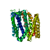
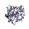
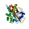
| Title | Crystal structure of substrate free cytochrome P450 NasF5053 from Streptomyces sp. NRRL F-5053 | |||||||||
 Components Components | cytochrome P450-F5053 | |||||||||
 Keywords Keywords | OXIDOREDUCTASE / biosynthetic protein / cytochrome P450 / cyclodipeptide / regio-selecitivty / stereo-selectivity / pyrroloindoline alkaloids / radical mediated reaction / F5053 / Streptomyces | |||||||||
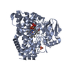
| Function / homology | BROMIDE ION / PROTOPORPHYRIN IX CONTAINING FE Function and homology information Function and homology information | |||||||||
| Biological species |  Streptomyces sp. NRRL F-5053 (bacteria) Streptomyces sp. NRRL F-5053 (bacteria) | |||||||||
| Method |  X-RAY DIFFRACTION / SYNCHROTRON / MOLECULAR REPLACEMENT / Resolution: 1.7 Å X-RAY DIFFRACTION / SYNCHROTRON / MOLECULAR REPLACEMENT / Resolution: 1.7 Å | |||||||||
 Authors Authors | Luo, Z. / Jia, X. / Sun, C. / Qu, X. / Kobe, B. | |||||||||
| Funding support |  Australia, Australia,  China, 2items China, 2items
| |||||||||
 Citation Citation | Journal: Nat Commun / Year: 2020 Title: Molecular basis of regio- and stereo-specificity in biosynthesis of bacterial heterodimeric diketopiperazines. Authors: Sun, C. / Luo, Z. / Zhang, W. / Tian, W. / Peng, H. / Lin, Z. / Deng, Z. / Kobe, B. / Jia, X. / Qu, X. | |||||||||
| History |
|
- Structure visualization
| Structure viewer | Molecule: MolmilJmol/JSmol |
|---|
- Downloads & links
-Download
| PDBx/mmCIF format | 6w0s.cif.gz | 317.5 KB | Display | PDBx/mmCIF format |
|---|---|---|---|---|
| PDB format | pdb6w0s.ent.gz | 253.9 KB | Display | PDB format |
| PDBx/mmJSON format | 6w0s.json.gz | Tree view | PDBx/mmJSON format | |
| Others |  Other downloads Other downloads |
-Validation report
| Arichive directory | https://data.pdbj.org/pub/pdb/validation_reports/w0/6w0sftp://data.pdbj.org/pub/pdb/validation_reports/w0/6w0s | HTTPS FTP |
|---|
-Related structure data
| Related structure data | 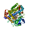 6vxvC 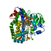 6vzaC 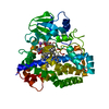 6vzbC 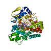 5wp2S S: Starting model for refinement C: citing same article ( |
|---|---|
| Similar structure data |


-Links
 PDBj
PDBj


- Assembly
| Deposited unit | 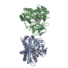
| ||||||||||||
|---|---|---|---|---|---|---|---|---|---|---|---|---|---|
| 1 | 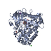
| ||||||||||||
| 2 | 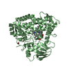
| ||||||||||||
| Unit cell |
|
-Components
-Protein , 1 types, 2 molecules AB
| #1: Protein | Mass: 43459.074 Da / Num. of mol.: 2 Source method: isolated from a genetically manipulated source Source: (gene. exp.) Streptomyces sp. NRRL F-5053 (bacteria)Production host: |
|---|
-Non-polymers , 6 types, 763 molecules 


| #2: Chemical | ChemComp-GOL /  Mass: 92.094 Da / Num. of mol.: 1 / Source method: obtained synthetically / Formula: C3H8O3 Mass: 92.094 Da / Num. of mol.: 1 / Source method: obtained synthetically / Formula: C3H8O3 | ||||||||
|---|---|---|---|---|---|---|---|---|---|
| #3: Chemical | ChemComp-BR /  Mass: 79.904 Da / Num. of mol.: 4 / Source method: obtained synthetically / Formula: Br Mass: 79.904 Da / Num. of mol.: 4 / Source method: obtained synthetically / Formula: Br#4: Chemical | ChemComp-CL /  Mass: 35.453 Da / Num. of mol.: 6 / Source method: obtained synthetically / Formula: Cl Mass: 35.453 Da / Num. of mol.: 6 / Source method: obtained synthetically / Formula: Cl#5: Chemical |  Mass: 22.990 Da / Num. of mol.: 2 / Source method: isolated from a natural source / Formula: Na Mass: 22.990 Da / Num. of mol.: 2 / Source method: isolated from a natural source / Formula: Na#6: Chemical |  Mass: 616.487 Da / Num. of mol.: 2 / Source method: isolated from a natural source / Formula: C34H32FeN4O4 / Feature type: SUBJECT OF INVESTIGATION Mass: 616.487 Da / Num. of mol.: 2 / Source method: isolated from a natural source / Formula: C34H32FeN4O4 / Feature type: SUBJECT OF INVESTIGATION#7: Water | ChemComp-HOH / | Mass: 18.015 Da / Num. of mol.: 748 / Source method: isolated from a natural source / Formula: H2O |
-Details
| Has ligand of interest | Y |
|---|
-Experimental details
-Experiment
| Experiment | Method: X-RAY DIFFRACTION / Number of used crystals: 1 |
|---|
- Sample preparation
| Crystal | Density Matthews: 2.17 Å3/Da / Density % sol: 43.3 % |
|---|---|
| Crystal grow | Temperature: 293 K / Method: vapor diffusion, hanging drop / Details: 0.2 M calcium chloride, 20% w/v PEG3350 |
-Data collection
| Diffraction | Mean temperature: 100 K / Serial crystal experiment: N |
|---|---|
| Diffraction source | Source: SYNCHROTRON / Site: Australian Synchrotron / Beamline: MX2 / Wavelength: 0.954 Å |
| Detector | Type: DECTRIS EIGER X 16M / Detector: PIXEL / Date: May 4, 2019 |
| Radiation | Protocol: SINGLE WAVELENGTH / Monochromatic (M) / Laue (L): M / Scattering type: x-ray |
| Radiation wavelength | Wavelength: 0.954 Å / Relative weight: 1 |
| Reflection | Resolution: 1.7→45.66 Å / Num. obs: 81192 / % possible obs: 99.8 % / Redundancy: 6.5 % / Biso Wilson estimate: 18.48 Å2 / CC1/2: 0.997 / Rmerge(I) obs: 0.108 / Rpim(I) all: 0.045 / Rrim(I) all: 0.117 / Χ2: 0.97 / Net I/σ(I): 9 |
| Reflection shell | Resolution: 1.7→1.73 Å / Rmerge(I) obs: 0.853 / Mean I/σ(I) obs: 2 / Num. unique obs: 4234 / CC1/2: 0.789 / Rpim(I) all: 0.392 / Rrim(I) all: 0.942 / Χ2: 1.04 |
- Processing
| Software |
| |||||||||||||||||||||||||||||||||||||||||||||||||||||||||||||||||||||||||||||||||||||||||||||||||||||||||||||||||||||||||||||||||||||||||||||||||||||||||||||||||||||||||||||||||||||||||||||||||||||||||||||||||||||||||
|---|---|---|---|---|---|---|---|---|---|---|---|---|---|---|---|---|---|---|---|---|---|---|---|---|---|---|---|---|---|---|---|---|---|---|---|---|---|---|---|---|---|---|---|---|---|---|---|---|---|---|---|---|---|---|---|---|---|---|---|---|---|---|---|---|---|---|---|---|---|---|---|---|---|---|---|---|---|---|---|---|---|---|---|---|---|---|---|---|---|---|---|---|---|---|---|---|---|---|---|---|---|---|---|---|---|---|---|---|---|---|---|---|---|---|---|---|---|---|---|---|---|---|---|---|---|---|---|---|---|---|---|---|---|---|---|---|---|---|---|---|---|---|---|---|---|---|---|---|---|---|---|---|---|---|---|---|---|---|---|---|---|---|---|---|---|---|---|---|---|---|---|---|---|---|---|---|---|---|---|---|---|---|---|---|---|---|---|---|---|---|---|---|---|---|---|---|---|---|---|---|---|---|---|---|---|---|---|---|---|---|---|---|---|---|---|---|---|---|
| Refinement | Method to determine structure: MOLECULAR REPLACEMENT Starting model: PDB entry 5WP2 Resolution: 1.7→43.06 Å / SU ML: 0.1794 / Cross valid method: FREE R-VALUE / σ(F): 1.34 / Phase error: 25.4871
| |||||||||||||||||||||||||||||||||||||||||||||||||||||||||||||||||||||||||||||||||||||||||||||||||||||||||||||||||||||||||||||||||||||||||||||||||||||||||||||||||||||||||||||||||||||||||||||||||||||||||||||||||||||||||
| Solvent computation | Shrinkage radii: 0.9 Å / VDW probe radii: 1.11 Å | |||||||||||||||||||||||||||||||||||||||||||||||||||||||||||||||||||||||||||||||||||||||||||||||||||||||||||||||||||||||||||||||||||||||||||||||||||||||||||||||||||||||||||||||||||||||||||||||||||||||||||||||||||||||||
| Displacement parameters | Biso mean: 24.07 Å2 | |||||||||||||||||||||||||||||||||||||||||||||||||||||||||||||||||||||||||||||||||||||||||||||||||||||||||||||||||||||||||||||||||||||||||||||||||||||||||||||||||||||||||||||||||||||||||||||||||||||||||||||||||||||||||
| Refinement step | Cycle: LAST / Resolution: 1.7→43.06 Å
| |||||||||||||||||||||||||||||||||||||||||||||||||||||||||||||||||||||||||||||||||||||||||||||||||||||||||||||||||||||||||||||||||||||||||||||||||||||||||||||||||||||||||||||||||||||||||||||||||||||||||||||||||||||||||
| Refine LS restraints |
| |||||||||||||||||||||||||||||||||||||||||||||||||||||||||||||||||||||||||||||||||||||||||||||||||||||||||||||||||||||||||||||||||||||||||||||||||||||||||||||||||||||||||||||||||||||||||||||||||||||||||||||||||||||||||
| LS refinement shell |
|
