[English] 日本語
 Yorodumi
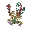
- PDB-6vzi: Crystal Structure of HIV-1 CAP256 RnS-3mut-2G-SOSIP.664 Prefusion... -
+ Open data
- Basic information
| Entry | Database: PDB / ID: 6vzi | |||||||||
|---|---|---|---|---|---|---|---|---|---|---|
| Title | Crystal Structure of HIV-1 CAP256 RnS-3mut-2G-SOSIP.664 Prefusion Env Trimer in Complex with Human Antibodies 3H109L and 35O22 at 3.5 Angstrom | |||||||||
 Components Components |
| |||||||||
 Keywords Keywords | VIRAL PROTEIN/IMMUNE SYSTEM / HIV-1 Envelope Prefusion Trimer / Entry Inhibitors / VIRAL PROTEIN / VIRAL PROTEIN-IMMUNE SYSTEM complex | |||||||||
| Function / homology |  Function and homology information Function and homology information: / positive regulation of establishment of T cell polarity / symbiont-mediated perturbation of host defense response / positive regulation of plasma membrane raft polarization / positive regulation of receptor clustering / host cell endosome membrane / clathrin-dependent endocytosis of virus by host cell / viral protein processing / fusion of virus membrane with host plasma membrane / fusion of virus membrane with host endosome membrane ...: / positive regulation of establishment of T cell polarity / symbiont-mediated perturbation of host defense response / positive regulation of plasma membrane raft polarization / positive regulation of receptor clustering / host cell endosome membrane / clathrin-dependent endocytosis of virus by host cell / viral protein processing / fusion of virus membrane with host plasma membrane / fusion of virus membrane with host endosome membrane / apoptotic process / viral envelope / virion attachment to host cell / host cell plasma membrane / virion membrane / structural molecule activity / membrane / plasma membrane Similarity search - Function | |||||||||
| Biological species |   Human immunodeficiency virus 1 Human immunodeficiency virus 1 Homo sapiens (human) Homo sapiens (human) | |||||||||
| Method |  X-RAY DIFFRACTION / X-RAY DIFFRACTION /  SYNCHROTRON / SYNCHROTRON /  MOLECULAR REPLACEMENT / Resolution: 2.716 Å MOLECULAR REPLACEMENT / Resolution: 2.716 Å | |||||||||
 Authors Authors | Lai, Y.-T. / Kwong, P.D. | |||||||||
 Citation Citation |  Journal: J.Virol. / Year: 2020 Journal: J.Virol. / Year: 2020Title: Development of a 3Mut-Apex-Stabilized Envelope Trimer That Expands HIV-1 Neutralization Breadth When Used To Boost Fusion Peptide-Directed Vaccine-Elicited Responses. Authors: Chuang, G.Y. / Lai, Y.T. / Boyington, J.C. / Cheng, C. / Geng, H. / Narpala, S. / Rawi, R. / Schmidt, S.D. / Tsybovsky, Y. / Verardi, R. / Xu, K. / Yang, Y. / Zhang, B. / Chambers, M. / ...Authors: Chuang, G.Y. / Lai, Y.T. / Boyington, J.C. / Cheng, C. / Geng, H. / Narpala, S. / Rawi, R. / Schmidt, S.D. / Tsybovsky, Y. / Verardi, R. / Xu, K. / Yang, Y. / Zhang, B. / Chambers, M. / Changela, A. / Corrigan, A.R. / Kong, R. / Olia, A.S. / Ou, L. / Sarfo, E.K. / Wang, S. / Wu, W. / Doria-Rose, N.A. / McDermott, A.B. / Mascola, J.R. / Kwong, P.D. | |||||||||
| History |
|
- Structure visualization
| Structure viewer | Molecule:  Molmil Molmil Jmol/JSmol Jmol/JSmol |
|---|
- Downloads & links
- Download
| PDBx/mmCIF format |  6vzi.cif.gz 6vzi.cif.gz | 269.9 KB | Display |  PDBx/mmCIF format PDBx/mmCIF format |
|---|---|---|---|---|
| PDB format |  pdb6vzi.ent.gz pdb6vzi.ent.gz | 211.6 KB | Display |  PDB format PDB format |
| PDBx/mmJSON format |  6vzi.json.gz 6vzi.json.gz | Tree view |  PDBx/mmJSON format PDBx/mmJSON format | |
| Others |  Other downloads Other downloads |
-Validation report
| Arichive directory |  https://data.pdbj.org/pub/pdb/validation_reports/vz/6vzi https://data.pdbj.org/pub/pdb/validation_reports/vz/6vzi ftp://data.pdbj.org/pub/pdb/validation_reports/vz/6vzi ftp://data.pdbj.org/pub/pdb/validation_reports/vz/6vzi | HTTPS FTP |
|---|
-Related structure data
| Related structure data |  6w03C  6mtjS C: citing same article ( S: Starting model for refinement |
|---|---|
| Similar structure data |
- Links
Links
- Assembly
| Deposited unit | 
| ||||||||
|---|---|---|---|---|---|---|---|---|---|
| 1 | 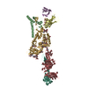
| ||||||||
| Unit cell |
|
- Components
-Envelope glycoprotein ... , 2 types, 2 molecules BG
| #1: Protein | Mass: 17324.734 Da / Num. of mol.: 1 / Fragment: ectodomain Mutation: I535N, I559P, T569G, I573F, K588E, D589V, T605C, Y609P, D636G, K651F, S655I Source method: isolated from a genetically manipulated source Source: (gene. exp.)   Human immunodeficiency virus 1 / Gene: env / Production host: Human immunodeficiency virus 1 / Gene: env / Production host:  Homo sapiens (human) / References: UniProt: W6ICH7 Homo sapiens (human) / References: UniProt: W6ICH7 |
|---|---|
| #4: Protein | Mass: 53421.621 Da / Num. of mol.: 1 / Mutation: A204I, N302M, T320L, A329P, S437P, E442N, A501C Source method: isolated from a genetically manipulated source Source: (gene. exp.)   Human immunodeficiency virus 1 / Gene: env / Production host: Human immunodeficiency virus 1 / Gene: env / Production host:  Homo sapiens (human) / References: UniProt: A0A0N9FF17 Homo sapiens (human) / References: UniProt: A0A0N9FF17 |
-Antibody , 4 types, 4 molecules DEHL
| #2: Antibody | Mass: 14663.281 Da / Num. of mol.: 1 / Mutation: E10T, L11T, K12T, A16S, I68N, K83T, F84S, Source method: isolated from a genetically manipulated source Source: (gene. exp.)  Homo sapiens (human) / Production host: Homo sapiens (human) / Production host:  Homo sapiens (human) Homo sapiens (human) |
|---|---|
| #3: Antibody | Mass: 12358.635 Da / Num. of mol.: 1 Source method: isolated from a genetically manipulated source Source: (gene. exp.)  Homo sapiens (human) / Production host: Homo sapiens (human) / Production host:  Homo sapiens (human) Homo sapiens (human) |
| #5: Antibody | Mass: 26255.498 Da / Num. of mol.: 1 Source method: isolated from a genetically manipulated source Source: (gene. exp.)  Homo sapiens (human) / Production host: Homo sapiens (human) / Production host:  Homo sapiens (human) Homo sapiens (human) |
| #6: Antibody | Mass: 23416.145 Da / Num. of mol.: 1 / Mutation: E184M, S188M Source method: isolated from a genetically manipulated source Source: (gene. exp.)  Homo sapiens (human) / Production host: Homo sapiens (human) / Production host:  Homo sapiens (human) Homo sapiens (human) |
-Sugars , 5 types, 15 molecules 
| #7: Polysaccharide | alpha-D-mannopyranose-(1-3)-alpha-D-mannopyranose-(1-6)-[alpha-D-mannopyranose-(1-3)]beta-D- ...alpha-D-mannopyranose-(1-3)-alpha-D-mannopyranose-(1-6)-[alpha-D-mannopyranose-(1-3)]beta-D-mannopyranose-(1-4)-2-acetamido-2-deoxy-beta-D-glucopyranose-(1-4)-2-acetamido-2-deoxy-beta-D-glucopyranose Source method: isolated from a genetically manipulated source | ||||||
|---|---|---|---|---|---|---|---|
| #8: Polysaccharide | Source method: isolated from a genetically manipulated source #9: Polysaccharide | Source method: isolated from a genetically manipulated source #10: Polysaccharide | alpha-D-mannopyranose-(1-2)-alpha-D-mannopyranose-(1-2)-alpha-D-mannopyranose-(1-3)-[alpha-D- ...alpha-D-mannopyranose-(1-2)-alpha-D-mannopyranose-(1-2)-alpha-D-mannopyranose-(1-3)-[alpha-D-mannopyranose-(1-2)-alpha-D-mannopyranose-(1-6)-[alpha-D-mannopyranose-(1-3)]alpha-D-mannopyranose-(1-6)]beta-D-mannopyranose-(1-4)-2-acetamido-2-deoxy-beta-D-glucopyranose-(1-4)-2-acetamido-2-deoxy-beta-D-glucopyranose | Source method: isolated from a genetically manipulated source #11: Sugar | ChemComp-NAG / |
-Details
| Has ligand of interest | N |
|---|---|
| Has protein modification | Y |
-Experimental details
-Experiment
| Experiment | Method:  X-RAY DIFFRACTION / Number of used crystals: 1 X-RAY DIFFRACTION / Number of used crystals: 1 |
|---|
- Sample preparation
| Crystal | Density Matthews: 5.53 Å3/Da / Density % sol: 77.77 % |
|---|---|
| Crystal grow | Temperature: 298 K / Method: evaporation Details: 60 mM sodium acetate pH 4.6, 120 mM ammonium sulfate and 6.3% PEG 4,000 |
-Data collection
| Diffraction | Mean temperature: 100 K / Serial crystal experiment: N | |||||||||||||||||||||||||||||||||||||||||||||||||||||||||||||||||||||||||||||||||||||||||||||||||||
|---|---|---|---|---|---|---|---|---|---|---|---|---|---|---|---|---|---|---|---|---|---|---|---|---|---|---|---|---|---|---|---|---|---|---|---|---|---|---|---|---|---|---|---|---|---|---|---|---|---|---|---|---|---|---|---|---|---|---|---|---|---|---|---|---|---|---|---|---|---|---|---|---|---|---|---|---|---|---|---|---|---|---|---|---|---|---|---|---|---|---|---|---|---|---|---|---|---|---|---|---|
| Diffraction source | Source:  SYNCHROTRON / Site: SYNCHROTRON / Site:  APS APS  / Beamline: 22-ID / Wavelength: 1 Å / Beamline: 22-ID / Wavelength: 1 Å | |||||||||||||||||||||||||||||||||||||||||||||||||||||||||||||||||||||||||||||||||||||||||||||||||||
| Detector | Type: RAYONIX MX300-HS / Detector: CCD / Date: Oct 13, 2018 | |||||||||||||||||||||||||||||||||||||||||||||||||||||||||||||||||||||||||||||||||||||||||||||||||||
| Radiation | Protocol: SINGLE WAVELENGTH / Monochromatic (M) / Laue (L): M / Scattering type: x-ray | |||||||||||||||||||||||||||||||||||||||||||||||||||||||||||||||||||||||||||||||||||||||||||||||||||
| Radiation wavelength | Wavelength: 1 Å / Relative weight: 1 | |||||||||||||||||||||||||||||||||||||||||||||||||||||||||||||||||||||||||||||||||||||||||||||||||||
| Reflection | Resolution: 2.7→50 Å / Num. obs: 53716 / % possible obs: 63.3 % / Redundancy: 7.1 % / Rmerge(I) obs: 0.128 / Rpim(I) all: 0.049 / Rrim(I) all: 0.138 / Χ2: 0.939 / Net I/σ(I): 5.5 / Num. measured all: 383837 | |||||||||||||||||||||||||||||||||||||||||||||||||||||||||||||||||||||||||||||||||||||||||||||||||||
| Reflection shell | Diffraction-ID: 1
|
- Processing
| Software |
| ||||||||||||||||||||||||||||||||||||||||||||||||||||||||||||||||||||||||
|---|---|---|---|---|---|---|---|---|---|---|---|---|---|---|---|---|---|---|---|---|---|---|---|---|---|---|---|---|---|---|---|---|---|---|---|---|---|---|---|---|---|---|---|---|---|---|---|---|---|---|---|---|---|---|---|---|---|---|---|---|---|---|---|---|---|---|---|---|---|---|---|---|---|
| Refinement | Method to determine structure:  MOLECULAR REPLACEMENT MOLECULAR REPLACEMENTStarting model: 6MTJ Resolution: 2.716→43.836 Å / SU ML: 0.41 / Cross valid method: THROUGHOUT / σ(F): 1.97 / Phase error: 28.12 / Stereochemistry target values: ML
| ||||||||||||||||||||||||||||||||||||||||||||||||||||||||||||||||||||||||
| Solvent computation | Shrinkage radii: 0.9 Å / VDW probe radii: 1.11 Å / Solvent model: FLAT BULK SOLVENT MODEL | ||||||||||||||||||||||||||||||||||||||||||||||||||||||||||||||||||||||||
| Displacement parameters | Biso max: 156.89 Å2 / Biso mean: 49.3208 Å2 / Biso min: 0.97 Å2 | ||||||||||||||||||||||||||||||||||||||||||||||||||||||||||||||||||||||||
| Refinement step | Cycle: final / Resolution: 2.716→43.836 Å
| ||||||||||||||||||||||||||||||||||||||||||||||||||||||||||||||||||||||||
| LS refinement shell | Refine-ID: X-RAY DIFFRACTION / Rfactor Rfree error: 0
|
 Movie
Movie Controller


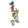

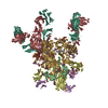
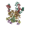

 PDBj
PDBj


