 Movie
Movie Controller

[English] 日本語
 Yorodumi
- PDB-6vwq: X-ray crystal structure of clavaminate synthase with vanadyl, suc... -
+ Open data

- Basic information
| Entry | Database: PDB / ID: 6vwq | ||||||
|---|---|---|---|---|---|---|---|
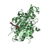
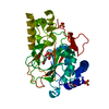
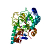
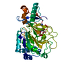
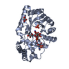
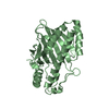
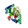
| Title | X-ray crystal structure of clavaminate synthase with vanadyl, succinate, and deoxyproclavaminic acid | ||||||
 Components Components | Clavaminate synthase 3 | ||||||
 Keywords Keywords | OXIDOREDUCTASE / iron / oxygenase / hydroxylase / oxacyclase / desaturase / alpha-ketoglutarate | ||||||
| Function / homology |  Function and homology information Function and homology informationantibiotic biosynthetic process / oxidoreductase activity / iron ion binding Similarity search - Function | ||||||
| Biological species |  Streptomyces antibioticus (bacteria) Streptomyces antibioticus (bacteria) | ||||||
| Method |  X-RAY DIFFRACTION / SYNCHROTRON / MOLECULAR REPLACEMENT / Resolution: 1.5 Å X-RAY DIFFRACTION / SYNCHROTRON / MOLECULAR REPLACEMENT / Resolution: 1.5 Å | ||||||
 Authors Authors | Boal, A.K. / Vavra, J. | ||||||
| Funding support |  United States, 1items United States, 1items
| ||||||
 Citation Citation | Journal: To Be Published Title: X-ray crystal structure of clavaminate synthase with vanadyl, succinate, and deoxyproclavaminic acid Authors: Vavra, J. / Dunham, N.P. / Krebs, C. / Boal, A.K. / Bollinger, J.M. | ||||||
| History |
|
- Structure visualization
| Structure viewer | Molecule: MolmilJmol/JSmol |
|---|
- Downloads & links
-Download
| PDBx/mmCIF format | 6vwq.cif.gz | 85.5 KB | Display | PDBx/mmCIF format |
|---|---|---|---|---|
| PDB format | pdb6vwq.ent.gz | 60.8 KB | Display | PDB format |
| PDBx/mmJSON format | 6vwq.json.gz | Tree view | PDBx/mmJSON format | |
| Others |  Other downloads Other downloads |
-Validation report
| Arichive directory | https://data.pdbj.org/pub/pdb/validation_reports/vw/6vwqftp://data.pdbj.org/pub/pdb/validation_reports/vw/6vwq | HTTPS FTP |
|---|
-Related structure data
| Related structure data | 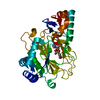 1drtS S: Starting model for refinement |
|---|---|
| Similar structure data |


-Links
 PDBj
PDBj- Assembly
| Deposited unit | 
| ||||||||||
|---|---|---|---|---|---|---|---|---|---|---|---|
| 1 |
| ||||||||||
| Unit cell |
|
-Components
| #1: Protein | Mass: 38370.152 Da / Num. of mol.: 1 Source method: isolated from a genetically manipulated source Source: (gene. exp.) Streptomyces antibioticus (bacteria) / Gene: cas3Production host: References: UniProt: I0CBY7, clavaminate synthase |
|---|---|
| #2: Chemical | ChemComp-VVO /   Mass: 66.941 Da / Num. of mol.: 1 / Source method: obtained synthetically / Formula: OV Mass: 66.941 Da / Num. of mol.: 1 / Source method: obtained synthetically / Formula: OV |
| #3: Chemical | ChemComp-SIN /   Mass: 118.088 Da / Num. of mol.: 1 / Source method: obtained synthetically / Formula: C4H6O4 Mass: 118.088 Da / Num. of mol.: 1 / Source method: obtained synthetically / Formula: C4H6O4 |
| #4: Chemical | ChemComp-RQJ /   Mass: 186.208 Da / Num. of mol.: 1 / Source method: obtained synthetically / Formula: C8H14N2O3 Mass: 186.208 Da / Num. of mol.: 1 / Source method: obtained synthetically / Formula: C8H14N2O3 |
| #5: Water | ChemComp-HOH /  Mass: 18.015 Da / Num. of mol.: 243 / Source method: isolated from a natural source / Formula: H2O Mass: 18.015 Da / Num. of mol.: 243 / Source method: isolated from a natural source / Formula: H2O |
| Has ligand of interest | Y |
-Experimental details
-Experiment
| Experiment | Method: X-RAY DIFFRACTION / Number of used crystals: 1 |
|---|
- Sample preparation
| Crystal | Density Matthews: 2.29 Å3/Da / Density % sol: 46.26 % |
|---|---|
| Crystal grow | Temperature: 298 K / Method: vapor diffusion, sitting drop / Details: magnesium acetate, PEG3350 |
-Data collection
| Diffraction | Mean temperature: 100 K / Serial crystal experiment: N |
|---|---|
| Diffraction source | Source: SYNCHROTRON / Site: APS / Beamline: 23-ID-B / Wavelength: 1.03314 Å |
| Detector | Type: DECTRIS EIGER X 16M / Detector: PIXEL / Date: Apr 7, 2019 |
| Radiation | Monochromator: Double crystal cryo-cooled Si(111) / Protocol: SINGLE WAVELENGTH / Monochromatic (M) / Laue (L): M / Scattering type: x-ray |
| Radiation wavelength | Wavelength: 1.03314 Å / Relative weight: 1 |
| Reflection | Resolution: 1.5→49.47 Å / Num. obs: 53621 / % possible obs: 99.3 % / Redundancy: 9.2 % / Biso Wilson estimate: 13.16 Å2 / Net I/σ(I): 18.38 |
| Reflection shell | Resolution: 1.5→1.53 Å |
- Processing
| Software |
| ||||||||||||||||||||||||
|---|---|---|---|---|---|---|---|---|---|---|---|---|---|---|---|---|---|---|---|---|---|---|---|---|---|
| Refinement | Method to determine structure: MOLECULAR REPLACEMENT Starting model: PDB entry 1DRT Resolution: 1.5→49.47 Å / SU ML: 0.136 / Cross valid method: FREE R-VALUE / σ(F): 1.37 / Phase error: 19.181 / Stereochemistry target values: GEOSTD + MONOMER LIBRARY
| ||||||||||||||||||||||||
| Solvent computation | Shrinkage radii: 0.9 Å / VDW probe radii: 1.11 Å / Solvent model: FLAT BULK SOLVENT MODEL | ||||||||||||||||||||||||
| Displacement parameters | Biso mean: 17.2 Å2 | ||||||||||||||||||||||||
| Refinement step | Cycle: LAST / Resolution: 1.5→49.47 Å
| ||||||||||||||||||||||||
| Refine LS restraints |
| ||||||||||||||||||||||||
| LS refinement shell | Highest resolution: 1.5 Å |
