 Movie
Movie Controller


+ Open data

- Basic information
| Entry | Database: PDB / ID: 6vu5 | |||||||||||||||||||||||||||||||||||||||||||||||||||
|---|---|---|---|---|---|---|---|---|---|---|---|---|---|---|---|---|---|---|---|---|---|---|---|---|---|---|---|---|---|---|---|---|---|---|---|---|---|---|---|---|---|---|---|---|---|---|---|---|---|---|---|---|
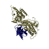
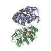
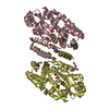
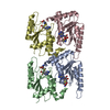
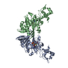
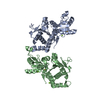
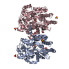
| Title | Structure of G-alpha-q bound to its chaperone Ric-8A | |||||||||||||||||||||||||||||||||||||||||||||||||||
 Components Components |
| |||||||||||||||||||||||||||||||||||||||||||||||||||
 Keywords Keywords | CHAPERONE / G protein alpha subunit / Ric-8 / molecular chaperone / G alpha folding / guanine nucleotide exchange factor (GEF) / cryoEM structure / protein complex / G protein-coupled receptor (GPCR) / phosphorylation / quality control. | |||||||||||||||||||||||||||||||||||||||||||||||||||
| Function / homology |  Function and homology information Function and homology informationcell-cell adhesion involved in gastrulation / cell migration involved in gastrulation / Fatty Acids bound to GPR40 (FFAR1) regulate insulin secretion / Acetylcholine regulates insulin secretion / sensory perception of itch / phospholipase C-activating G protein-coupled glutamate receptor signaling pathway / basement membrane organization / PLC beta mediated events / phospholipase C-activating serotonin receptor signaling pathway / regulation of platelet activation ...cell-cell adhesion involved in gastrulation / cell migration involved in gastrulation / Fatty Acids bound to GPR40 (FFAR1) regulate insulin secretion / Acetylcholine regulates insulin secretion / sensory perception of itch / phospholipase C-activating G protein-coupled glutamate receptor signaling pathway / basement membrane organization / PLC beta mediated events / phospholipase C-activating serotonin receptor signaling pathway / regulation of platelet activation / entrainment of circadian clock / vasculature development / regulation of canonical Wnt signaling pathway / glutamate receptor signaling pathway / mast cell degranulation / phototransduction, visible light / response to light stimulus / photoreceptor outer segment / G-protein alpha-subunit binding / postsynaptic cytosol / cellular response to acidic pH / hormone-mediated signaling pathway / gastrulation / protein folding chaperone / guanyl-nucleotide exchange factor activity / Turbulent (oscillatory, disturbed) flow shear stress activates signaling by PIEZO1 and integrins in endothelial cells / GTPase activator activity / neuropeptide signaling pathway / response to prostaglandin E / visual learning / G protein-coupled receptor binding / G-protein beta/gamma-subunit complex binding / adenylate cyclase-inhibiting G protein-coupled receptor signaling pathway / blood coagulation / G protein-coupled acetylcholine receptor signaling pathway / Thromboxane signalling through TP receptor / G-protein activation / ADP signalling through P2Y purinoceptor 1 / Cooperation of PDCL (PhLP1) and TRiC/CCT in G-protein beta folding / heterotrimeric G-protein complex / Thrombin signalling through proteinase activated receptors (PARs) / adenylate cyclase-activating G protein-coupled receptor signaling pathway / G protein activity / cell cortex / nuclear membrane / High laminar flow shear stress activates signaling by PIEZO1 and PECAM1:CDH5:KDR in endothelial cells / phospholipase C-activating G protein-coupled receptor signaling pathway / G alpha (q) signalling events / in utero embryonic development / Hydrolases; Acting on acid anhydrides; Acting on GTP to facilitate cellular and subcellular movement / protein stabilization / G protein-coupled receptor signaling pathway / lysosomal membrane / GTPase activity / GTP binding / Golgi apparatus / extracellular exosome / membrane / metal ion binding / plasma membrane / cytoplasm Similarity search - Function | |||||||||||||||||||||||||||||||||||||||||||||||||||
| Biological species |   Homo sapiens (human) Homo sapiens (human) | |||||||||||||||||||||||||||||||||||||||||||||||||||
| Method | ELECTRON MICROSCOPY / single particle reconstruction / cryo EM / Resolution: 3.5 Å | |||||||||||||||||||||||||||||||||||||||||||||||||||
 Authors Authors | Seven, A.B. / Hilger, D. | |||||||||||||||||||||||||||||||||||||||||||||||||||
| Funding support |  United States, 1items United States, 1items
| |||||||||||||||||||||||||||||||||||||||||||||||||||
 Citation Citation | Journal: Cell Rep / Year: 2020 Title: Structures of Gα Proteins in Complex with Their Chaperone Reveal Quality Control Mechanisms. Authors: Alpay Burak Seven / Daniel Hilger / Makaía M Papasergi-Scott / Li Zhang / Qianhui Qu / Brian K Kobilka / Gregory G Tall / Georgios Skiniotis / Abstract: Many chaperones promote nascent polypeptide folding followed by substrate release through ATP-dependent conformational changes. Here we show cryoEM structures of Gα subunit folding intermediates in ...Many chaperones promote nascent polypeptide folding followed by substrate release through ATP-dependent conformational changes. Here we show cryoEM structures of Gα subunit folding intermediates in complex with full-length Ric-8A, a unique chaperone-client system in which substrate release is facilitated by guanine nucleotide binding to the client G protein. The structures of Ric-8A-Gα and Ric-8A-Gα complexes reveal that the chaperone employs its extended C-terminal region to cradle the Ras-like domain of Gα, positioning the Ras core in contact with the Ric-8A core while engaging its switch2 nucleotide binding region. The C-terminal α5 helix of Gα is held away from the Ras-like domain through Ric-8A core domain interactions, which critically depend on recognition of the Gα C terminus by the chaperone. The structures, complemented with biochemical and cellular chaperoning data, support a folding quality control mechanism that ensures proper formation of the C-terminal α5 helix before allowing GTP-gated release of Gα from Ric-8A. | |||||||||||||||||||||||||||||||||||||||||||||||||||
| History |
|
- Structure visualization
| Movie |
Movie viewer |
|---|---|
| Structure viewer | Molecule: MolmilJmol/JSmol |
- Downloads & links
-Download
| PDBx/mmCIF format | 6vu5.cif.gz | 121.6 KB | Display | PDBx/mmCIF format |
|---|---|---|---|---|
| PDB format | pdb6vu5.ent.gz | 88.5 KB | Display | PDB format |
| PDBx/mmJSON format | 6vu5.json.gz | Tree view | PDBx/mmJSON format | |
| Others |  Other downloads Other downloads |
-Validation report
| Arichive directory | https://data.pdbj.org/pub/pdb/validation_reports/vu/6vu5ftp://data.pdbj.org/pub/pdb/validation_reports/vu/6vu5 | HTTPS FTP |
|---|
-Related structure data
| Related structure data | 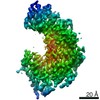 21387MC  6vu8C M: map data used to model this data C: citing same article ( |
|---|---|
| Similar structure data |


-Links
 PDBj
PDBj


- Assembly
| Deposited unit | 
|
|---|---|
| 1 |
|
-Components
| #1: Protein | Mass: 53908.895 Da / Num. of mol.: 1 Source method: isolated from a genetically manipulated source Source: (gene. exp.)  Trichoplusia ni (cabbage looper) / References: UniProt: Q80ZG1*PLUS Trichoplusia ni (cabbage looper) / References: UniProt: Q80ZG1*PLUS |
|---|---|
| #2: Protein | Mass: 42197.027 Da / Num. of mol.: 1 Source method: isolated from a genetically manipulated source Source: (gene. exp.) Homo sapiens (human) / Gene: GNAQ, GAQ / Production host: Trichoplusia ni (cabbage looper) / References: UniProt: P50148 |
| Has ligand of interest | N |
| Has protein modification | Y |
| Sequence details | The full sequence of Ric-8A is QGEFMEPRAVADALETGEEDAVTEALRSFNREHSQSFTFDDAQQEDRKRLAKLLVSVLE ...The full sequence of Ric-8A is QGEFMEPRAV |
-Experimental details
-Experiment
| Experiment | Method: ELECTRON MICROSCOPY |
|---|---|
| EM experiment | Aggregation state: PARTICLE / 3D reconstruction method: single particle reconstruction |
- Sample preparation
| Component | Name: Complex of Ric-8A with G alpha q / Type: COMPLEX / Entity ID: all / Source: RECOMBINANT |
|---|---|
| Source (natural) | Organism: |
| Source (recombinant) | Organism: Trichoplusia ni (cabbage looper) |
| Buffer solution | pH: 7.5 |
| Specimen | Embedding applied: NO / Shadowing applied: NO / Staining applied: NO / Vitrification applied: YES |
| Vitrification | Cryogen name: ETHANE |
- Electron microscopy imaging
| Experimental equipment |  Model: Titan Krios / Image courtesy: FEI Company |
|---|---|
| Microscopy | Model: FEI TITAN KRIOS |
| Electron gun | Electron source:  FIELD EMISSION GUN / Accelerating voltage: 300 kV / Illumination mode: FLOOD BEAM FIELD EMISSION GUN / Accelerating voltage: 300 kV / Illumination mode: FLOOD BEAM |
| Electron lens | Mode: BRIGHT FIELD / Nominal defocus max: 2000 nm / Nominal defocus min: 1000 nm / Cs: 2.7 mm / C2 aperture diameter: 50 µm |
| Image recording | Electron dose: 1.3 e/Å2 / Detector mode: COUNTING / Film or detector model: GATAN K2 QUANTUM (4k x 4k) |
- Processing
| Software | Name: PHENIX / Version: 1.17.1_3660: / Classification: refinement | ||||||||||||||||||||||||
|---|---|---|---|---|---|---|---|---|---|---|---|---|---|---|---|---|---|---|---|---|---|---|---|---|---|
| EM software |
| ||||||||||||||||||||||||
| CTF correction | Type: PHASE FLIPPING AND AMPLITUDE CORRECTION | ||||||||||||||||||||||||
| Symmetry | Point symmetry: C1 (asymmetric) | ||||||||||||||||||||||||
| 3D reconstruction | Resolution: 3.5 Å / Resolution method: FSC 0.143 CUT-OFF / Num. of particles: 70439 / Symmetry type: POINT | ||||||||||||||||||||||||
| Refine LS restraints |
|
