[English] 日本語
Yorodumi
- EMDB-21387: Structure of G-alpha-q bound to its chaperone Ric-8A -

+
Open data


ID or keywords:

Loading...

-
Basic information

Entry
Database: EMDB / ID: EMD-21387
TitleStructure of G-alpha-q bound to its chaperone Ric-8A
Map dataRelion-Local-Res-Job229
Sample
  • Complex: Complex of Ric-8A with G alpha q
    • Protein or peptide: Resistance to inhibitors of cholinesterase-8A (Ric-8A)
    • Protein or peptide: Guanine nucleotide-binding protein G(q) subunit alpha
Function / homology
Function and homology information


cell-cell adhesion involved in gastrulation / cell migration involved in gastrulation / Fatty Acids bound to GPR40 (FFAR1) regulate insulin secretion / Acetylcholine regulates insulin secretion / basement membrane organization / PLC beta mediated events / phospholipase C-activating dopamine receptor signaling pathway / entrainment of circadian clock / regulation of platelet activation / phototransduction, visible light ...cell-cell adhesion involved in gastrulation / cell migration involved in gastrulation / Fatty Acids bound to GPR40 (FFAR1) regulate insulin secretion / Acetylcholine regulates insulin secretion / basement membrane organization / PLC beta mediated events / phospholipase C-activating dopamine receptor signaling pathway / entrainment of circadian clock / regulation of platelet activation / phototransduction, visible light / vasculature development / glutamate receptor signaling pathway / regulation of canonical Wnt signaling pathway / action potential / activation of phospholipase C activity / response to light stimulus / photoreceptor outer segment / gastrulation / G-protein alpha-subunit binding / adenylate cyclase-inhibiting G protein-coupled receptor signaling pathway / GTPase activator activity / guanyl-nucleotide exchange factor activity / G protein-coupled receptor binding / G-protein beta/gamma-subunit complex binding / negative regulation of protein kinase activity / Thromboxane signalling through TP receptor / visual learning / G-protein activation / G protein-coupled acetylcholine receptor signaling pathway / adenylate cyclase-activating G protein-coupled receptor signaling pathway / ADP signalling through P2Y purinoceptor 1 / Cooperation of PDCL (PhLP1) and TRiC/CCT in G-protein beta folding / heterotrimeric G-protein complex / blood coagulation / Thrombin signalling through proteinase activated receptors (PARs) / G alpha (q) signalling events / nuclear membrane / in utero embryonic development / protein stabilization / G protein-coupled receptor signaling pathway / lysosomal membrane / GTPase activity / synapse / GTP binding / Golgi apparatus / extracellular exosome / membrane / metal ion binding / plasma membrane / cytoplasm
Similarity search - Function
Synembryn / Guanine nucleotide exchange factor, Ric8 / Guanine nucleotide exchange factor synembryn / G-protein alpha subunit, group Q / G-alpha domain profile. / Guanine nucleotide binding protein (G-protein), alpha subunit / G protein alpha subunit, helical insertion / G-protein alpha subunit / G protein alpha subunit / Armadillo-like helical ...Synembryn / Guanine nucleotide exchange factor, Ric8 / Guanine nucleotide exchange factor synembryn / G-protein alpha subunit, group Q / G-alpha domain profile. / Guanine nucleotide binding protein (G-protein), alpha subunit / G protein alpha subunit, helical insertion / G-protein alpha subunit / G protein alpha subunit / Armadillo-like helical / Armadillo-type fold / P-loop containing nucleoside triphosphate hydrolase
Similarity search - Domain/homology
Guanine nucleotide-binding protein G(q) subunit alpha / Synembryn-A
Similarity search - Component
Biological speciesRattus norvegicus/Homo sapiens (Norway rat) / Rattus norvegicus (Norway rat) / Homo sapiens (human)
Methodsingle particle reconstruction / cryo EM / Resolution: 3.5 Å
AuthorsSeven AB / Hilger D
Funding support United States, 1 items
OrganizationGrant numberCountry
National Institutes of Health/National Institute of Neurological Disorders and Stroke (NIH/NINDS)NS092695 United States
Citation
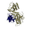
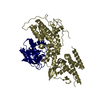
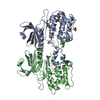
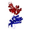
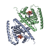
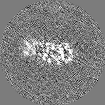
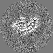
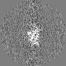
Journal: Cell Rep / Year: 2020
Title: Structures of Gα Proteins in Complex with Their Chaperone Reveal Quality Control Mechanisms.
Authors: Alpay Burak Seven / Daniel Hilger / Makaía M Papasergi-Scott / Li Zhang / Qianhui Qu / Brian K Kobilka / Gregory G Tall / Georgios Skiniotis /
Abstract: Many chaperones promote nascent polypeptide folding followed by substrate release through ATP-dependent conformational changes. Here we show cryoEM structures of Gα subunit folding intermediates in ...Many chaperones promote nascent polypeptide folding followed by substrate release through ATP-dependent conformational changes. Here we show cryoEM structures of Gα subunit folding intermediates in complex with full-length Ric-8A, a unique chaperone-client system in which substrate release is facilitated by guanine nucleotide binding to the client G protein. The structures of Ric-8A-Gα and Ric-8A-Gα complexes reveal that the chaperone employs its extended C-terminal region to cradle the Ras-like domain of Gα, positioning the Ras core in contact with the Ric-8A core while engaging its switch2 nucleotide binding region. The C-terminal α5 helix of Gα is held away from the Ras-like domain through Ric-8A core domain interactions, which critically depend on recognition of the Gα C terminus by the chaperone. The structures, complemented with biochemical and cellular chaperoning data, support a folding quality control mechanism that ensures proper formation of the C-terminal α5 helix before allowing GTP-gated release of Gα from Ric-8A.
History
DepositionFeb 14, 2020-
Header (metadata) releaseFeb 26, 2020-
Map releaseMar 18, 2020-
UpdateDec 2, 2020-
Current statusDec 2, 2020Processing site: RCSB / Status: Released

-
Structure visualization
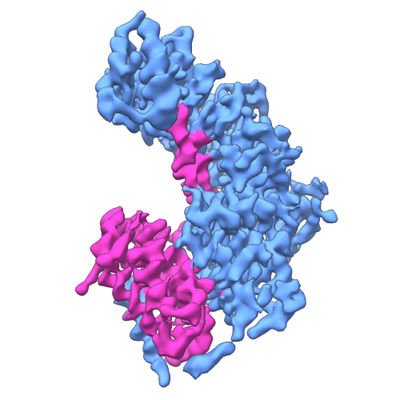
Movie
  • Surface view with section colored by density value
  • Surface level: 0.0367
  • Imaged by UCSF Chimera
  • Download
  • Surface view colored by radius
  • Surface level: 0.0367
  • Imaged by UCSF Chimera
  • Download
  • Surface view with fitted model
  • Atomic models: PDB-6vu5
  • Surface level: 0.0367
  • Imaged by UCSF Chimera
  • Download
Movie viewer
Structure viewerEM map:
SurfViewMolmilJmol/JSmol
Supplemental images

Downloads & links

-
Map

FileDownload / File: emd_21387.map.gz / Format: CCP4 / Size: 38.4 MB / Type: IMAGE STORED AS FLOATING POINT NUMBER (4 BYTES)
AnnotationRelion-Local-Res-Job229
Voxel sizeX=Y=Z: 0.82 Å
Density
Contour LevelBy AUTHOR: 0.0367 / Movie #1: 0.0367
Minimum - Maximum-0.16094479 - 0.25380152
Average (Standard dev.)-0.00002226122 (±0.0061857146)
SymmetrySpace group: 1
Details

EMDB XML:

Map geometry
Axis orderXYZ
Origin000
Dimensions216216216
Spacing216216216
CellA=B=C: 177.12 Å
α=β=γ: 90.0 °

CCP4 map header:

modeImage stored as Reals
Å/pix. X/Y/Z0.820.820.82
M x/y/z216216216
origin x/y/z0.0000.0000.000
length x/y/z177.120177.120177.120
α/β/γ90.00090.00090.000
MAP C/R/S123
start NC/NR/NS000
NC/NR/NS216216216
D min/max/mean-0.1610.254-0.000

-
Supplemental data

-
Half map: #2

Fileemd_21387_half_map_1.map
Projections & Slices
AxesZYX

Projections

Slices (1/2)
Density Histograms

-
Half map: #1

Fileemd_21387_half_map_2.map
Projections & Slices
AxesZYX

Projections

Slices (1/2)
Density Histograms

-
Sample components

-
Entire : Complex of Ric-8A with G alpha q

EntireName: Complex of Ric-8A with G alpha q
Components
  • Complex: Complex of Ric-8A with G alpha q
    • Protein or peptide: Resistance to inhibitors of cholinesterase-8A (Ric-8A)
    • Protein or peptide: Guanine nucleotide-binding protein G(q) subunit alpha

-
Supramolecule #1: Complex of Ric-8A with G alpha q

SupramoleculeName: Complex of Ric-8A with G alpha q / type: complex / ID: 1 / Parent: 0 / Macromolecule list: all
Source (natural)Organism: Rattus norvegicus/Homo sapiens (Norway rat)
Recombinant expressionOrganism: Trichoplusia ni (cabbage looper)

-
Macromolecule #1: Resistance to inhibitors of cholinesterase-8A (Ric-8A)

MacromoleculeName: Resistance to inhibitors of cholinesterase-8A (Ric-8A)
type: protein_or_peptide / ID: 1 / Number of copies: 1 / Enantiomer: LEVO
Source (natural)Organism: Rattus norvegicus (Norway rat)
Molecular weightTheoretical: 53.908895 KDa
Recombinant expressionOrganism: Trichoplusia ni (cabbage looper)
SequenceString: QGEFMEPRAV ADALETGEED AVTEALRSFN REHSQSFTFD DAQQEDRKRL AKLLVSVLEQ GLSPKHRVTW LQTIRILSRD RSCLDSFAS RQSLHALACY ADIAISEEPI PQPPDMDVLL ESLKCLCNLV LSSPTAQMLA AEARLVVRLA ERVGLYRKRS Y PHEVQFFD ...String:
QGEFMEPRAV ADALETGEED AVTEALRSFN REHSQSFTFD DAQQEDRKRL AKLLVSVLEQ GLSPKHRVTW LQTIRILSRD RSCLDSFAS RQSLHALACY ADIAISEEPI PQPPDMDVLL ESLKCLCNLV LSSPTAQMLA AEARLVVRLA ERVGLYRKRS Y PHEVQFFD LRLLFLLTAL RTDVRQQLFQ ELHGVRLLTD ALELTLGVAP KENPLVILPA QETERAMEIL KVLFNITFDS VK REVDEED AALYRYLGTL LRHCVMADAA GDRTEEFHGH TVNLLGNLPL KCLDVLLALE LHEGSLEFMG VNMDVINALL AFL EKRLHQ THRLKECVAP VLSVLTECAR MHRPARKFLK AQVLPPLRDV RTRPEVGDLL RNKLVRLMTH LDTDVKRVAA EFLF VLCSE SVPRFIKYTG YGNAAGLLAA RGLMAGGRPE GQY(SEP)EDED(TPO)D TEEYREAKAS (UNK)(UNK)(UNK) (UNK)(UNK)(UNK)(UNK)(UNK)(UNK) (UNK)(UNK)(UNK)(UNK)(UNK)(UNK)(UNK)(UNK)(UNK)(UNK) (UNK)(UNK)(UNK)(UNK)(UNK)(UNK)(UNK)(UNK)

-
Macromolecule #2: Guanine nucleotide-binding protein G(q) subunit alpha

MacromoleculeName: Guanine nucleotide-binding protein G(q) subunit alpha / type: protein_or_peptide / ID: 2 / Number of copies: 1 / Enantiomer: LEVO
Source (natural)Organism: Homo sapiens (human)
Molecular weightTheoretical: 42.197027 KDa
Recombinant expressionOrganism: Trichoplusia ni (cabbage looper)
SequenceString: MTLESIMACC LSEEAKEARR INDEIERQLR RDKRDARREL KLLLLGTGES GKSTFIKQMR IIHGSGYSDE DKRGFTKLVY QNIFTAMQA MIRAMDTLKI PYKYEHNKAH AQLVREVDVE KVSAFENPYV DAIKSLWNDP GIQECYDRRR EYQLSDSTKY Y LNDLDRVA ...String:
MTLESIMACC LSEEAKEARR INDEIERQLR RDKRDARREL KLLLLGTGES GKSTFIKQMR IIHGSGYSDE DKRGFTKLVY QNIFTAMQA MIRAMDTLKI PYKYEHNKAH AQLVREVDVE KVSAFENPYV DAIKSLWNDP GIQECYDRRR EYQLSDSTKY Y LNDLDRVA DPAYLPTQQD VLRVRVPTTG IIEYPFDLQS VIFRMVDVGG QRSERRKWIH CFENVTSIMF LVALSEYDQV LV ESDNENR MEESKALFRT IITYPWFQNS SVILFLNKKD LLEEKIMYSH LVDYFPEYDG PQRDAQAARE FILKMFVDLN PDS DKIIYS HFTCATDTEN IRFVFAAVKD TILQLNLKEY NLV

-
Experimental details

-
Structure determination

Methodcryo EM
Processingsingle particle reconstruction
Aggregation stateparticle

-
Sample preparation

BufferpH: 7.5
VitrificationCryogen name: ETHANE

-
Electron microscopy

MicroscopeFEI TITAN KRIOS
Electron beamAcceleration voltage: 300 kV / Electron source: FIELD EMISSION GUN
Electron opticsC2 aperture diameter: 50.0 µm / Illumination mode: FLOOD BEAM / Imaging mode: BRIGHT FIELDBright-field microscopy / Cs: 2.7 mm / Nominal defocus max: 2.0 µm / Nominal defocus min: 1.0 µm
Image recordingFilm or detector model: GATAN K2 QUANTUM (4k x 4k) / Detector mode: COUNTING / Average electron dose: 1.3 e/Å2
Experimental equipment
Model: Titan Krios / Image courtesy: FEI Company

-
Image processing

Initial angle assignmentType: OTHER
Final angle assignmentType: OTHER
Final reconstructionApplied symmetry - Point group: C1 (asymmetric) / Resolution.type: BY AUTHOR / Resolution: 3.5 Å / Resolution method: FSC 0.143 CUT-OFF / Software - Name: RELION (ver. 3.0) / Number images used: 70439

+
About Yorodumi

-
News

-
Feb 9, 2022. New format data for meta-information of EMDB entries

New format data for meta-information of EMDB entries

  • Version 3 of the EMDB header file is now the official format.
  • The previous official version 1.9 will be removed from the archive.

Related info.:EMDB header

External links:wwPDB to switch to version 3 of the EMDB data model

-
Aug 12, 2020. Covid-19 info

Covid-19 info

URL: https://pdbj.org/emnavi/covid19.php

New page: Covid-19 featured information page in EM Navigator.

Related info.:Covid-19 info / Mar 5, 2020. Novel coronavirus structure data

+
Mar 5, 2020. Novel coronavirus structure data

Novel coronavirus structure data

Related info.:Yorodumi Speices / Aug 12, 2020. Covid-19 info

External links:COVID-19 featured content - PDBj / Molecule of the Month (242):Coronavirus Proteases

+
Jan 31, 2019. EMDB accession codes are about to change! (news from PDBe EMDB page)

EMDB accession codes are about to change! (news from PDBe EMDB page)

  • The allocation of 4 digits for EMDB accession codes will soon come to an end. Whilst these codes will remain in use, new EMDB accession codes will include an additional digit and will expand incrementally as the available range of codes is exhausted. The current 4-digit format prefixed with “EMD-” (i.e. EMD-XXXX) will advance to a 5-digit format (i.e. EMD-XXXXX), and so on. It is currently estimated that the 4-digit codes will be depleted around Spring 2019, at which point the 5-digit format will come into force.
  • The EM Navigator/Yorodumi systems omit the EMD- prefix.

Related info.:Q: What is EMD? / ID/Accession-code notation in Yorodumi/EM Navigator

External links:EMDB Accession Codes are Changing Soon! / Contact to PDBj

+
Jul 12, 2017. Major update of PDB

Major update of PDB

  • wwPDB released updated PDB data conforming to the new PDBx/mmCIF dictionary.
  • This is a major update changing the version number from 4 to 5, and with Remediation, in which all the entries are updated.
  • In this update, many items about electron microscopy experimental information are reorganized (e.g. em_software).
  • Now, EM Navigator and Yorodumi are based on the updated data.

External links:wwPDB Remediation / Enriched Model Files Conforming to OneDep Data Standards Now Available in the PDB FTP Archive

-
Yorodumi

Thousand views of thousand structures

  • Yorodumi is a browser for structure data from EMDB, PDB, SASBDB, etc.
  • This page is also the successor to EM Navigator detail page, and also detail information page/front-end page for Omokage search.
  • The word "yorodu" (or yorozu) is an old Japanese word meaning "ten thousand". "mi" (miru) is to see.

Related info.:EMDB / PDB / SASBDB / Comparison of 3 databanks / Yorodumi Search / Aug 31, 2016. New EM Navigator & Yorodumi / Yorodumi Papers / Jmol/JSmol / Function and homology information / Changes in new EM Navigator and Yorodumi

Read more