 Movie
Movie Controller

[English] 日本語
 Yorodumi
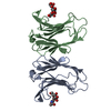
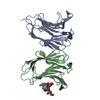
- PDB-6vto: Crystal structure of human Galectin-7 in complex with 4-O-beta-D-... -
+ Open data

- Basic information
| Entry | Database: PDB / ID: 6vto | |||||||||||||||||||||
|---|---|---|---|---|---|---|---|---|---|---|---|---|---|---|---|---|---|---|---|---|---|---|
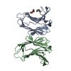
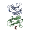
| Title | Crystal structure of human Galectin-7 in complex with 4-O-beta-D-Galactopyranosyl-D-glucose | |||||||||||||||||||||
 Components Components | Galectin-7 | |||||||||||||||||||||
 Keywords Keywords | SUGAR BINDING PROTEIN / Human Galectin-7 / lactose-furanose | |||||||||||||||||||||
| Function / homology |  Function and homology information Function and homology informationDifferentiation of Keratinocytes in Interfollicular Epidermis in Mammalian Skin / heterophilic cell-cell adhesion / carbohydrate binding / apoptotic process / : / extracellular exosome / nucleus / cytoplasm Similarity search - Function | |||||||||||||||||||||
| Biological species |  Homo sapiens (human) Homo sapiens (human) | |||||||||||||||||||||
| Method |  X-RAY DIFFRACTION / SYNCHROTRON / MOLECULAR REPLACEMENT / molecular replacement / Resolution: 1.69 Å X-RAY DIFFRACTION / SYNCHROTRON / MOLECULAR REPLACEMENT / molecular replacement / Resolution: 1.69 Å | |||||||||||||||||||||
 Authors Authors | Pham, N.T.H. / Calmettes, C. / Doucet, N. | |||||||||||||||||||||
| Funding support |  Canada, Canada,  United States, 6items United States, 6items
| |||||||||||||||||||||
 Citation Citation | Journal: J.Biol.Chem. / Year: 2021 Title: Perturbing dimer interactions and allosteric communication modulates the immunosuppressive activity of human galectin-7. Authors: Pham, N.T.H. / Letourneau, M. / Fortier, M. / Begin, G. / Al-Abdul-Wahid, M.S. / Pucci, F. / Folch, B. / Rooman, M. / Chatenet, D. / St-Pierre, Y. / Lague, P. / Calmettes, C. / Doucet, N. | |||||||||||||||||||||
| History |
|
- Structure visualization
| Structure viewer | Molecule: MolmilJmol/JSmol |
|---|
- Downloads & links
-Download
| PDBx/mmCIF format | 6vto.cif.gz | 121.8 KB | Display | PDBx/mmCIF format |
|---|---|---|---|---|
| PDB format | pdb6vto.ent.gz | 94.2 KB | Display | PDB format |
| PDBx/mmJSON format | 6vto.json.gz | Tree view | PDBx/mmJSON format | |
| Others |  Other downloads Other downloads |
-Validation report
| Arichive directory | https://data.pdbj.org/pub/pdb/validation_reports/vt/6vtoftp://data.pdbj.org/pub/pdb/validation_reports/vt/6vto | HTTPS FTP |
|---|
-Related structure data
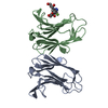
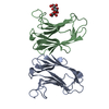
| Related structure data |  6vtpC 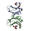 6vtqC 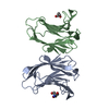 6vtrC 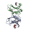 6vtsC  4galS S: Starting model for refinement C: citing same article ( |
|---|---|
| Similar structure data |


-Links
 PDBj
PDBj- Assembly
| Deposited unit | 
| ||||||||
|---|---|---|---|---|---|---|---|---|---|
| 1 |
| ||||||||
| Unit cell |
|
-Components
| #1: Protein | Mass: 14965.850 Da / Num. of mol.: 2 Source method: isolated from a genetically manipulated source Source: (gene. exp.) Homo sapiens (human) / Gene: LGALS7, PIG1, LGALS7B / Plasmid: pET22b / Production host:  #2: Chemical | 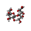  Mass: 342.296 Da / Num. of mol.: 2 / Source method: obtained synthetically / Formula: C12H22O11 / Feature type: SUBJECT OF INVESTIGATION Mass: 342.296 Da / Num. of mol.: 2 / Source method: obtained synthetically / Formula: C12H22O11 / Feature type: SUBJECT OF INVESTIGATION#3: Water | ChemComp-HOH / |  Mass: 18.015 Da / Num. of mol.: 313 / Source method: isolated from a natural source / Formula: H2O Mass: 18.015 Da / Num. of mol.: 313 / Source method: isolated from a natural source / Formula: H2OHas ligand of interest | Y | |
|---|
-Experimental details
-Experiment
| Experiment | Method: X-RAY DIFFRACTION / Number of used crystals: 1 |
|---|
- Sample preparation
| Crystal | Density Matthews: 2.19 Å3/Da / Density % sol: 43.94 % |
|---|---|
| Crystal grow | Temperature: 295 K / Method: vapor diffusion, sitting drop / pH: 8 Details: 0.1 M Sodium Chloride, 0.1 M Tris pH 8, 20 % PEG3350, 5 % Glycerol Temp details: Room temperature |
-Data collection
| Diffraction | Mean temperature: 100 K / Serial crystal experiment: N |
|---|---|
| Diffraction source | Source: SYNCHROTRON / Site: CLSI / Beamline: 08B1-1 / Wavelength: 1.0332 Å |
| Detector | Type: RAYONIX MX300HE / Detector: CCD / Date: Feb 18, 2019 / Details: 16 tiled fiber-optic tapers |
| Radiation | Monochromator: KOHZU double crystal monochromator (DCM) / Protocol: SINGLE WAVELENGTH / Monochromatic (M) / Laue (L): M / Scattering type: x-ray |
| Radiation wavelength | Wavelength: 1.0332 Å / Relative weight: 1 |
| Reflection | Resolution: 1.69→45.19 Å / Num. obs: 30152 / % possible obs: 99.11 % / Redundancy: 6.85 % / Biso Wilson estimate: 30.173 Å2 / CC1/2: 0.999 / Rmerge(I) obs: 0.086 / Rrim(I) all: 0.093 / Net I/σ(I): 15.7 / Num. measured all: 206559 |
| Reflection shell | Resolution: 1.69→1.75 Å / Redundancy: 7.17 % / Rmerge(I) obs: 0.836 / Mean I/σ(I) obs: 3 / Num. measured obs: 21014 / Num. unique obs: 2930 / CC1/2: 0.839 / Rrim(I) all: 0.901 / % possible all: 99.25 |
-Phasing
| Phasing | Method: molecular replacement | |||||||||
|---|---|---|---|---|---|---|---|---|---|---|
| Phasing MR |
|
- Processing
| Software |
| |||||||||||||||||||||||||||||||||||||||||||||||||||||||||||||||
|---|---|---|---|---|---|---|---|---|---|---|---|---|---|---|---|---|---|---|---|---|---|---|---|---|---|---|---|---|---|---|---|---|---|---|---|---|---|---|---|---|---|---|---|---|---|---|---|---|---|---|---|---|---|---|---|---|---|---|---|---|---|---|---|---|
| Refinement | Method to determine structure: MOLECULAR REPLACEMENT Starting model: 4gal Resolution: 1.69→45.19 Å / SU ML: 0.23 / Cross valid method: THROUGHOUT / σ(F): 1.34 / Phase error: 22.29 / Stereochemistry target values: ML
| |||||||||||||||||||||||||||||||||||||||||||||||||||||||||||||||
| Solvent computation | Shrinkage radii: 0.9 Å / VDW probe radii: 1.11 Å / Solvent model: FLAT BULK SOLVENT MODEL | |||||||||||||||||||||||||||||||||||||||||||||||||||||||||||||||
| Displacement parameters | Biso max: 95.98 Å2 / Biso mean: 21.037 Å2 / Biso min: 5.25 Å2 | |||||||||||||||||||||||||||||||||||||||||||||||||||||||||||||||
| Refinement step | Cycle: final / Resolution: 1.69→45.19 Å
| |||||||||||||||||||||||||||||||||||||||||||||||||||||||||||||||
| LS refinement shell | Refine-ID: X-RAY DIFFRACTION / Rfactor Rfree error: 0 / Total num. of bins used: 8
|
