 Movie
Movie Controller

[English] 日本語
 Yorodumi
- PDB-6vqj: Mammalian V-ATPase from rat brain collar and peripheral stalks ro... -
+ Open data

- Basic information
| Entry | Database: PDB / ID: 6vqj | ||||||
|---|---|---|---|---|---|---|---|
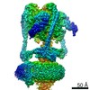
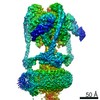
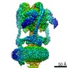
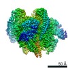
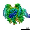
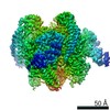
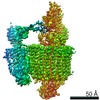
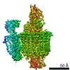
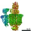
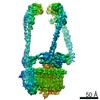
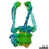
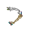
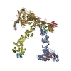
| Title | Mammalian V-ATPase from rat brain collar and peripheral stalks rotational state 2 (from focused refinement) | ||||||
 Components Components |
| ||||||
 Keywords Keywords | PROTON TRANSPORT / membrane protein complex / rotary atpase | ||||||
| Function / homology |  Function and homology information Function and homology informationIon channel transport / Transferrin endocytosis and recycling / Amino acids regulate mTORC1 / Insulin receptor recycling / proton-transporting V-type ATPase, V1 domain / synaptic vesicle lumen acidification / P-type proton-exporting transporter activity / extrinsic component of synaptic vesicle membrane / vacuolar proton-transporting V-type ATPase, V1 domain / vacuolar proton-transporting V-type ATPase, V0 domain ...Ion channel transport / Transferrin endocytosis and recycling / Amino acids regulate mTORC1 / Insulin receptor recycling / proton-transporting V-type ATPase, V1 domain / synaptic vesicle lumen acidification / P-type proton-exporting transporter activity / extrinsic component of synaptic vesicle membrane / vacuolar proton-transporting V-type ATPase, V1 domain / vacuolar proton-transporting V-type ATPase, V0 domain / clathrin-coated vesicle membrane / proton-transporting V-type ATPase complex / vacuolar proton-transporting V-type ATPase complex / vacuolar acidification / ROS and RNS production in phagocytes / Neutrophil degranulation / microvillus / proton-transporting ATPase activity, rotational mechanism / regulation of macroautophagy / proton transmembrane transport / apical part of cell / terminal bouton / melanosome / synaptic vesicle membrane / ATPase binding / endosome / apical plasma membrane / perinuclear region of cytoplasm / ATP hydrolysis activity / membrane / plasma membrane / cytosol / cytoplasm Similarity search - Function | ||||||
| Biological species |  | ||||||
| Method | ELECTRON MICROSCOPY / single particle reconstruction / cryo EM / Resolution: 5.7 Å | ||||||
 Authors Authors | Abbas, Y.M. / Rubinstein, J.L. | ||||||
| Funding support |  Canada, 1items Canada, 1items
| ||||||
 Citation Citation | Journal: Science / Year: 2020 Title: Structure of V-ATPase from the mammalian brain. Authors: Yazan M Abbas / Di Wu / Stephanie A Bueler / Carol V Robinson / John L Rubinstein /  Abstract: In neurons, the loading of neurotransmitters into synaptic vesicles uses energy from proton-pumping vesicular- or vacuolar-type adenosine triphosphatases (V-ATPases). These membrane protein complexes ...In neurons, the loading of neurotransmitters into synaptic vesicles uses energy from proton-pumping vesicular- or vacuolar-type adenosine triphosphatases (V-ATPases). These membrane protein complexes possess numerous subunit isoforms, which complicates their analysis. We isolated homogeneous rat brain V-ATPase through its interaction with SidK, a effector protein. Cryo-electron microscopy allowed the construction of an atomic model, defining the enzyme's ATP:proton ratio as 3:10 and revealing a homolog of yeast subunit f in the membrane region, which we tentatively identify as RNAseK. The c ring encloses the transmembrane anchors for cleaved ATP6AP1/Ac45 and ATP6AP2/PRR, the latter of which is the (pro)renin receptor that, in other contexts, is involved in both Wnt signaling and the renin-angiotensin system that regulates blood pressure. This structure shows how ATP6AP1/Ac45 and ATP6AP2/PRR enable assembly of the enzyme's catalytic and membrane regions. | ||||||
| History |
|
- Structure visualization
| Movie |
Movie viewer |
|---|---|
| Structure viewer | Molecule: MolmilJmol/JSmol |
- Downloads & links
-Download
| PDBx/mmCIF format | 6vqj.cif.gz | 179 KB | Display | PDBx/mmCIF format |
|---|---|---|---|---|
| PDB format | pdb6vqj.ent.gz | 111.9 KB | Display | PDB format |
| PDBx/mmJSON format | 6vqj.json.gz | Tree view | PDBx/mmJSON format | |
| Others |  Other downloads Other downloads |
-Validation report
| Arichive directory | https://data.pdbj.org/pub/pdb/validation_reports/vq/6vqjftp://data.pdbj.org/pub/pdb/validation_reports/vq/6vqj | HTTPS FTP |
|---|
-Related structure data
| Related structure data | 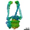 21352MC 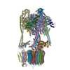 6vq6C 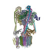 6vq7C 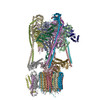 6vq8C 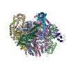 6vq9C 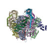 6vqaC 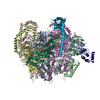 6vqbC 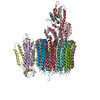 6vqcC 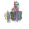 6vqgC 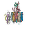 6vqhC 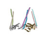 6vqiC 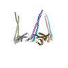 6vqkC M: map data used to model this data C: citing same article ( |
|---|---|
| Similar structure data |

-Links
 PDBj
PDBj


- Assembly
| Deposited unit | 
|
|---|---|
| 1 |
|
-Components
| #1: Protein | Mass: 43958.453 Da / Num. of mol.: 1 / Source method: isolated from a natural source / Source: (natural) | ||||
|---|---|---|---|---|---|
| #2: Protein | Mass: 26167.453 Da / Num. of mol.: 3 / Source method: isolated from a natural source / Source: (natural) #3: Protein | Mass: 13690.476 Da / Num. of mol.: 3 / Source method: isolated from a natural source / Source: (natural) #4: Protein | | Mass: 96429.438 Da / Num. of mol.: 1 / Source method: isolated from a natural source / Source: (natural) |
-Experimental details
-Experiment
| Experiment | Method: ELECTRON MICROSCOPY |
|---|---|
| EM experiment | Aggregation state: PARTICLE / 3D reconstruction method: single particle reconstruction |
- Sample preparation
| Component | Name: Peripheral stalk and collar region of mammalian V-ATPase composed of subunits E1, G2, N-terminal domain of a1, and C1. Type: COMPLEX / Entity ID: all / Source: NATURAL |
|---|---|
| Source (natural) | Organism: |
| Buffer solution | pH: 7 |
| Specimen | Embedding applied: NO / Shadowing applied: NO / Staining applied: NO / Vitrification applied: YES |
| Vitrification | Cryogen name: ETHANE-PROPANE |
- Electron microscopy imaging
| Experimental equipment |  Model: Titan Krios / Image courtesy: FEI Company |
|---|---|
| Microscopy | Model: FEI TITAN KRIOS |
| Electron gun | Electron source:  FIELD EMISSION GUN / Accelerating voltage: 300 kV / Illumination mode: FLOOD BEAM FIELD EMISSION GUN / Accelerating voltage: 300 kV / Illumination mode: FLOOD BEAM |
| Electron lens | Mode: BRIGHT FIELD |
| Image recording | Electron dose: 43 e/Å2 / Film or detector model: FEI FALCON III (4k x 4k) |
- Processing
| EM software | Name: cryoSPARC / Category: 3D reconstruction |
|---|---|
| CTF correction | Type: PHASE FLIPPING AND AMPLITUDE CORRECTION |
| Symmetry | Point symmetry: C1 (asymmetric) |
| 3D reconstruction | Resolution: 5.7 Å / Resolution method: FSC 0.143 CUT-OFF / Num. of particles: 74789 / Algorithm: BACK PROJECTION / Symmetry type: POINT |
