 Movie
Movie Controller

[English] 日本語
 Yorodumi
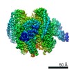
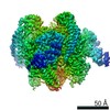
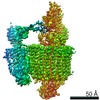
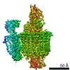
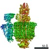
- EMDB-21317: Mammalian V-ATPase from rat brain with the Legionella pneumophila... -
+ Open data

- Basic information
| Entry | Database: EMDB / ID: EMD-21317 | |||||||||
|---|---|---|---|---|---|---|---|---|---|---|
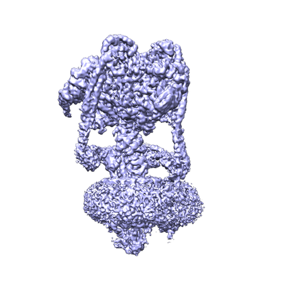
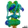
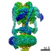
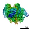
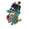
| Title | Mammalian V-ATPase from rat brain with the Legionella pneumophila effector protein SidK - rotational state 1 non-uniform refinement | |||||||||
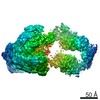
 Map data Map data | Mammalian rat brain V-ATPase with SidK bound, non-uniform refinement conformational state 1 | |||||||||
 Sample Sample |
| |||||||||
| Function / homology |  Function and homology information Function and homology informationMetabolism of Angiotensinogen to Angiotensins / Ion channel transport / Transferrin endocytosis and recycling / Amino acids regulate mTORC1 / symbiont-mediated suppression of host phagosome acidification / Insulin receptor recycling / RHOA GTPase cycle / negative regulation of autophagic cell death / eye pigmentation / central nervous system maturation ...Metabolism of Angiotensinogen to Angiotensins / Ion channel transport / Transferrin endocytosis and recycling / Amino acids regulate mTORC1 / symbiont-mediated suppression of host phagosome acidification / Insulin receptor recycling / RHOA GTPase cycle / negative regulation of autophagic cell death / eye pigmentation / central nervous system maturation / proton-transporting V-type ATPase, V1 domain / positive regulation of transforming growth factor beta1 production / rostrocaudal neural tube patterning / synaptic vesicle lumen acidification / P-type proton-exporting transporter activity / proton-transporting V-type ATPase, V0 domain / cellular response to increased oxygen levels / extrinsic component of synaptic vesicle membrane / vacuolar proton-transporting V-type ATPase, V1 domain / endosome to plasma membrane protein transport / vacuolar proton-transporting V-type ATPase, V0 domain / lysosomal lumen acidification / clathrin-coated vesicle membrane / endosomal lumen acidification / proton-transporting V-type ATPase complex / head morphogenesis / protein localization to cilium / vacuolar proton-transporting V-type ATPase complex / osteoclast development / vacuolar acidification / : / ROS and RNS production in phagocytes / dendritic spine membrane / Neutrophil degranulation / ATPase complex / homeostatic process / microvillus / ATPase activator activity / regulation of MAPK cascade / autophagosome membrane / proton-transporting ATPase activity, rotational mechanism / cilium assembly / regulation of macroautophagy / transporter activator activity / H+-transporting two-sector ATPase / positive regulation of Wnt signaling pathway / ATP metabolic process / angiotensin maturation / receptor-mediated endocytosis of virus by host cell / ruffle / RNA endonuclease activity / endoplasmic reticulum-Golgi intermediate compartment membrane / receptor-mediated endocytosis / proton transmembrane transport / transmembrane transport / small GTPase binding / apical part of cell / terminal bouton / synaptic vesicle / melanosome / positive regulation of canonical Wnt signaling pathway / synaptic vesicle membrane / ATPase binding / signaling receptor activity / cell body / intracellular iron ion homeostasis / positive regulation of ERK1 and ERK2 cascade / lysosome / endosome / endosome membrane / apical plasma membrane / nuclear speck / cilium / external side of plasma membrane / axon / lysosomal membrane / ubiquitin protein ligase binding / centrosome / endoplasmic reticulum membrane / perinuclear region of cytoplasm / Golgi apparatus / ATP hydrolysis activity / : / nucleoplasm / ATP binding / membrane / plasma membrane / cytoplasm / cytosol Similarity search - Function | |||||||||
| Biological species |  | |||||||||
| Method | single particle reconstruction / cryo EM / Resolution: 3.9 Å | |||||||||
 Authors Authors | Abbas YM / Rubinstein JL | |||||||||
| Funding support |  Canada, 1 items Canada, 1 items
| |||||||||
 Citation Citation | Journal: Science / Year: 2020 Title: Structure of V-ATPase from the mammalian brain. Authors: Yazan M Abbas / Di Wu / Stephanie A Bueler / Carol V Robinson / John L Rubinstein /  Abstract: In neurons, the loading of neurotransmitters into synaptic vesicles uses energy from proton-pumping vesicular- or vacuolar-type adenosine triphosphatases (V-ATPases). These membrane protein complexes ...In neurons, the loading of neurotransmitters into synaptic vesicles uses energy from proton-pumping vesicular- or vacuolar-type adenosine triphosphatases (V-ATPases). These membrane protein complexes possess numerous subunit isoforms, which complicates their analysis. We isolated homogeneous rat brain V-ATPase through its interaction with SidK, a effector protein. Cryo-electron microscopy allowed the construction of an atomic model, defining the enzyme's ATP:proton ratio as 3:10 and revealing a homolog of yeast subunit f in the membrane region, which we tentatively identify as RNAseK. The c ring encloses the transmembrane anchors for cleaved ATP6AP1/Ac45 and ATP6AP2/PRR, the latter of which is the (pro)renin receptor that, in other contexts, is involved in both Wnt signaling and the renin-angiotensin system that regulates blood pressure. This structure shows how ATP6AP1/Ac45 and ATP6AP2/PRR enable assembly of the enzyme's catalytic and membrane regions. | |||||||||
| History |
|
- Structure visualization
| Movie |
Movie viewer |
|---|---|
| Structure viewer | EM map: SurfViewMolmilJmol/JSmol |
| Supplemental images |
- Downloads & links
-EMDB archive
| Map data | emd_21317.map.gz | 141.6 MB | EMDB map data format | |
|---|---|---|---|---|
| Header (meta data) | emd-21317-v30.xmlemd-21317.xml | 9.1 KB 9.1 KB | Display Display | EMDB header |
| Images |  emd_21317.png emd_21317.png | 113.4 KB | ||
| Archive directory |  http://ftp.pdbj.org/pub/emdb/structures/EMD-21317ftp://ftp.pdbj.org/pub/emdb/structures/EMD-21317 http://ftp.pdbj.org/pub/emdb/structures/EMD-21317ftp://ftp.pdbj.org/pub/emdb/structures/EMD-21317 | HTTPS FTP |
-Related structure data
| Related structure data | 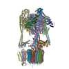 6vq6MC 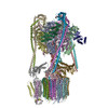 6vq7C 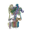 6vq8C 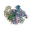 6vq9C 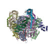 6vqaC 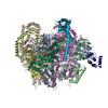 6vqbC 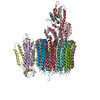 6vqcC 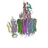 6vqgC 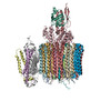 6vqhC  6vqiC  6vqjC  6vqkC M: atomic model generated by this map C: citing same article ( |
|---|---|
| Similar structure data |


-Links
| EMDB pages | EMDB (EBI/PDBe) / EMDataResource |
|---|---|
| Related items in Molecule of the Month |


-Map
| File | Download / File: emd_21317.map.gz / Format: CCP4 / Size: 149.9 MB / Type: IMAGE STORED AS FLOATING POINT NUMBER (4 BYTES) | ||||||||||||||||||||||||||||||||||||||||||||||||||||||||||||||||||||
|---|---|---|---|---|---|---|---|---|---|---|---|---|---|---|---|---|---|---|---|---|---|---|---|---|---|---|---|---|---|---|---|---|---|---|---|---|---|---|---|---|---|---|---|---|---|---|---|---|---|---|---|---|---|---|---|---|---|---|---|---|---|---|---|---|---|---|---|---|---|
| Annotation | Mammalian rat brain V-ATPase with SidK bound, non-uniform refinement conformational state 1 | ||||||||||||||||||||||||||||||||||||||||||||||||||||||||||||||||||||
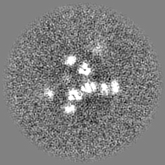
| Projections & slices | Image control
Images are generated by Spider. | ||||||||||||||||||||||||||||||||||||||||||||||||||||||||||||||||||||
| Voxel size | X=Y=Z: 1.06 Å | ||||||||||||||||||||||||||||||||||||||||||||||||||||||||||||||||||||
| Density |
| ||||||||||||||||||||||||||||||||||||||||||||||||||||||||||||||||||||
| Symmetry | Space group: 1 | ||||||||||||||||||||||||||||||||||||||||||||||||||||||||||||||||||||
| Details | EMDB XML:
CCP4 map header:
| ||||||||||||||||||||||||||||||||||||||||||||||||||||||||||||||||||||
 Z (Sec.)
 Y (Row.)
 X (Col.)


-Supplemental data
- Sample components
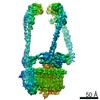
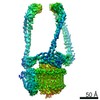
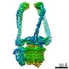
-Entire : Mammalian rat brain V-ATPase collar and peripheral stalks state 1...
| Entire | Name: Mammalian rat brain V-ATPase collar and peripheral stalks state 1 - from focused refinement |
|---|---|
| Components |
|
-Supramolecule #1: Mammalian rat brain V-ATPase collar and peripheral stalks state 1...
| Supramolecule | Name: Mammalian rat brain V-ATPase collar and peripheral stalks state 1 - from focused refinement type: complex / ID: 1 / Parent: 0 / Macromolecule list: #1-#6 |
|---|---|
| Source (natural) | Organism: |
-Experimental details
-Structure determination
| Method | cryo EM |
|---|---|
 Processing Processing | single particle reconstruction |
| Aggregation state | particle |
-Sample preparation
| Buffer | pH: 7 |
|---|---|
| Vitrification | Cryogen name: ETHANE-PROPANE |
- Electron microscopy
| Microscope | FEI TITAN KRIOS |
|---|---|
| Image recording | Film or detector model: FEI FALCON III (4k x 4k) / Average electron dose: 43.0 e/Å2 |
| Electron beam | Acceleration voltage: 300 kV / Electron source:  FIELD EMISSION GUN FIELD EMISSION GUN |
| Electron optics | Illumination mode: FLOOD BEAM / Imaging mode: BRIGHT FIELD |
| Experimental equipment |  Model: Titan Krios / Image courtesy: FEI Company |
-Image processing
| Final reconstruction | Applied symmetry - Point group: C1 (asymmetric) / Algorithm: BACK PROJECTION / Resolution.type: BY AUTHOR / Resolution: 3.9 Å / Resolution method: FSC 0.143 CUT-OFF / Software - Name: cryoSPARC / Number images used: 90648 |
|---|---|
| Initial angle assignment | Type: RANDOM ASSIGNMENT |
| Final angle assignment | Type: PROJECTION MATCHING |
