 Movie
Movie Controller


+ Open data

- Basic information
| Entry | Database: PDB / ID: 6vnx | ||||||
|---|---|---|---|---|---|---|---|
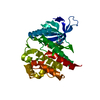
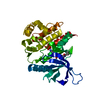
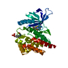
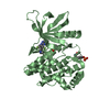
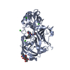
| Title | Crystal structure of TYK2 kinase with compound 19 | ||||||
 Components Components | Non-receptor tyrosine-protein kinase TYK2 | ||||||
 Keywords Keywords | TRANSFERASE/INHIBITOR / kinase / TRANSFERASE / TRANSFERASE-INHIBITOR complex | ||||||
| Function / homology |  Function and homology information Function and homology informationtype III interferon-mediated signaling pathway / interleukin-10-mediated signaling pathway / interleukin-12 receptor complex / interleukin-23 receptor complex / Interleukin-23 signaling / positive regulation of T-helper 17 type immune response / interleukin-12-mediated signaling pathway / positive regulation of NK T cell proliferation / interleukin-23-mediated signaling pathway / Interleukin-12 signaling ...type III interferon-mediated signaling pathway / interleukin-10-mediated signaling pathway / interleukin-12 receptor complex / interleukin-23 receptor complex / Interleukin-23 signaling / positive regulation of T-helper 17 type immune response / interleukin-12-mediated signaling pathway / positive regulation of NK T cell proliferation / interleukin-23-mediated signaling pathway / Interleukin-12 signaling / IL-6-type cytokine receptor ligand interactions / Interleukin-27 signaling / Interleukin-35 Signalling / growth hormone receptor binding / Other interleukin signaling / extrinsic component of plasma membrane / Interleukin-20 family signaling / extrinsic component of cytoplasmic side of plasma membrane / Interleukin-6 signaling / type I interferon-mediated signaling pathway / MAPK3 (ERK1) activation / MAPK1 (ERK2) activation / Interleukin-10 signaling / positive regulation of interleukin-17 production / Regulation of IFNA/IFNB signaling / positive regulation of natural killer cell proliferation / growth hormone receptor signaling pathway via JAK-STAT / type II interferon-mediated signaling pathway / cell surface receptor signaling pathway via JAK-STAT / Signaling by CSF3 (G-CSF) / positive regulation of T cell proliferation / positive regulation of receptor signaling pathway via JAK-STAT / non-specific protein-tyrosine kinase / non-membrane spanning protein tyrosine kinase activity / cellular response to virus / positive regulation of protein localization to nucleus / Inactivation of CSF3 (G-CSF) signaling / Evasion by RSV of host interferon responses / cytoplasmic side of plasma membrane / cytokine-mediated signaling pathway / positive regulation of type II interferon production / Interferon alpha/beta signaling / Signaling by ALK fusions and activated point mutants / protein tyrosine kinase activity / Interleukin-4 and Interleukin-13 signaling / Potential therapeutics for SARS / protein phosphorylation / cell differentiation / cell population proliferation / signaling receptor complex / intracellular signal transduction / immune response / SARS-CoV-2 activates/modulates innate and adaptive immune responses / extracellular exosome / ATP binding / nucleus / plasma membrane / cytosol / cytoplasm Similarity search - Function | ||||||
| Biological species |  Homo sapiens (human) Homo sapiens (human) | ||||||
| Method |  X-RAY DIFFRACTION / SYNCHROTRON / MOLECULAR REPLACEMENT / Resolution: 2.18 Å X-RAY DIFFRACTION / SYNCHROTRON / MOLECULAR REPLACEMENT / Resolution: 2.18 Å | ||||||
 Authors Authors | Vajdos, F.F. | ||||||
 Citation Citation | Journal: Bioorg.Med.Chem. / Year: 2020 Title: Design and optimization of a series of 4-(3-azabicyclo[3.1.0]hexan-3-yl)pyrimidin-2-amines: Dual inhibitors of TYK2 and JAK1. Authors: Fensome, A. / Ambler, C.M. / Arnold, E. / Banker, M.E. / Clark, J.D. / Dowty, M.E. / Efremov, I.V. / Flick, A. / Gerstenberger, B.S. / Gifford, R.S. / Gopalsamy, A. / Hegen, M. / Jussif, J. ...Authors: Fensome, A. / Ambler, C.M. / Arnold, E. / Banker, M.E. / Clark, J.D. / Dowty, M.E. / Efremov, I.V. / Flick, A. / Gerstenberger, B.S. / Gifford, R.S. / Gopalsamy, A. / Hegen, M. / Jussif, J. / Limburg, D.C. / Lin, T.H. / Pierce, B.S. / Sharma, R. / Trujillo, J.I. / Vajdos, F.F. / Vincent, F. / Wan, Z.K. / Xing, L. / Yang, X. / Yang, X. | ||||||
| History |
|
- Structure visualization
| Structure viewer | Molecule: MolmilJmol/JSmol |
|---|
- Downloads & links
-Download
| PDBx/mmCIF format | 6vnx.cif.gz | 80.1 KB | Display | PDBx/mmCIF format |
|---|---|---|---|---|
| PDB format | pdb6vnx.ent.gz | 56.5 KB | Display | PDB format |
| PDBx/mmJSON format | 6vnx.json.gz | Tree view | PDBx/mmJSON format | |
| Others |  Other downloads Other downloads |
-Validation report
| Arichive directory | https://data.pdbj.org/pub/pdb/validation_reports/vn/6vnxftp://data.pdbj.org/pub/pdb/validation_reports/vn/6vnx | HTTPS FTP |
|---|
-Related structure data
| Related structure data |  6vnsC  6vnvC  6vnyC  6w8lC 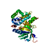 3lxpS S: Starting model for refinement C: citing same article ( |
|---|---|
| Similar structure data |


-Links
 PDBj
PDBj


- Assembly
| Deposited unit | 
| ||||||||
|---|---|---|---|---|---|---|---|---|---|
| 1 |
| ||||||||
| Unit cell |
|
-Components
| #1: Protein | Mass: 36550.570 Da / Num. of mol.: 1 / Fragment: kinase domain / Mutation: C936A, C1142A, Q969A, E971A, K972A Source method: isolated from a genetically manipulated source Source: (gene. exp.) Homo sapiens (human) / Gene: TYK2 / Cell line (production host): Sf21 / Production host:   Spodoptera frugiperda (fall armyworm) Spodoptera frugiperda (fall armyworm)References: UniProt: P29597, non-specific protein-tyrosine kinase |
|---|---|
| #2: Chemical | ChemComp-R4V / (  Mass: 407.393 Da / Num. of mol.: 1 / Source method: obtained synthetically / Formula: C18H20F3N7O / Feature type: SUBJECT OF INVESTIGATION Mass: 407.393 Da / Num. of mol.: 1 / Source method: obtained synthetically / Formula: C18H20F3N7O / Feature type: SUBJECT OF INVESTIGATION |
| #3: Water | ChemComp-HOH /  Mass: 18.015 Da / Num. of mol.: 125 / Source method: isolated from a natural source / Formula: H2O Mass: 18.015 Da / Num. of mol.: 125 / Source method: isolated from a natural source / Formula: H2O |
| Has ligand of interest | Y |
| Has protein modification | Y |
-Experimental details
-Experiment
| Experiment | Method: X-RAY DIFFRACTION / Number of used crystals: 1 |
|---|
- Sample preparation
| Crystal | Density Matthews: 1.9 Å3/Da / Density % sol: 35.32 % / Mosaicity: 0 ° |
|---|---|
| Crystal grow | Temperature: 298 K / Method: evaporation / pH: 8 Details: 0.1 M bis-tris pH 5.5, 0.25 M NaCl, 10 mM TCEP, 27-33% PEG-3350 |
-Data collection
| Diffraction | Mean temperature: 100 K / Serial crystal experiment: N | |||||||||||||||||||||||||||||||||||||||||||||||||||||||||||||||||||||||||||||||||||||||||||||||||||||||||||||||||||||||||
|---|---|---|---|---|---|---|---|---|---|---|---|---|---|---|---|---|---|---|---|---|---|---|---|---|---|---|---|---|---|---|---|---|---|---|---|---|---|---|---|---|---|---|---|---|---|---|---|---|---|---|---|---|---|---|---|---|---|---|---|---|---|---|---|---|---|---|---|---|---|---|---|---|---|---|---|---|---|---|---|---|---|---|---|---|---|---|---|---|---|---|---|---|---|---|---|---|---|---|---|---|---|---|---|---|---|---|---|---|---|---|---|---|---|---|---|---|---|---|---|---|---|---|
| Diffraction source | Source: SYNCHROTRON / Site: APS  / Beamline: 17-ID / Wavelength: 1 Å / Beamline: 17-ID / Wavelength: 1 Å | |||||||||||||||||||||||||||||||||||||||||||||||||||||||||||||||||||||||||||||||||||||||||||||||||||||||||||||||||||||||||
| Detector | Type: DECTRIS PILATUS 6M / Detector: PIXEL / Date: Feb 27, 2013 | |||||||||||||||||||||||||||||||||||||||||||||||||||||||||||||||||||||||||||||||||||||||||||||||||||||||||||||||||||||||||
| Radiation | Protocol: SINGLE WAVELENGTH / Monochromatic (M) / Laue (L): M / Scattering type: x-ray | |||||||||||||||||||||||||||||||||||||||||||||||||||||||||||||||||||||||||||||||||||||||||||||||||||||||||||||||||||||||||
| Radiation wavelength | Wavelength: 1 Å / Relative weight: 1 | |||||||||||||||||||||||||||||||||||||||||||||||||||||||||||||||||||||||||||||||||||||||||||||||||||||||||||||||||||||||||
| Reflection | Resolution: 2.179→103.825 Å / Num. all: 14813 / Num. obs: 14813 / % possible obs: 97.3 % / Redundancy: 5.3 % / Biso Wilson estimate: 32.55 Å2 / Rpim(I) all: 0.045 / Rrim(I) all: 0.108 / Rsym value: 0.088 / Net I/av σ(I): 7.2 / Net I/σ(I): 12.7 / Num. measured all: 78551 | |||||||||||||||||||||||||||||||||||||||||||||||||||||||||||||||||||||||||||||||||||||||||||||||||||||||||||||||||||||||||
| Reflection shell | Diffraction-ID: 1
|
- Processing
| Software |
| ||||||||||||||||||||||||||||||||||||||||||||||||||||||||||||||||||||||||||||||||||||||||||||||||||||||||||||
|---|---|---|---|---|---|---|---|---|---|---|---|---|---|---|---|---|---|---|---|---|---|---|---|---|---|---|---|---|---|---|---|---|---|---|---|---|---|---|---|---|---|---|---|---|---|---|---|---|---|---|---|---|---|---|---|---|---|---|---|---|---|---|---|---|---|---|---|---|---|---|---|---|---|---|---|---|---|---|---|---|---|---|---|---|---|---|---|---|---|---|---|---|---|---|---|---|---|---|---|---|---|---|---|---|---|---|---|---|---|
| Refinement | Method to determine structure: MOLECULAR REPLACEMENT Starting model: 3LXP Resolution: 2.18→51.91 Å / Cor.coef. Fo:Fc: 0.9136 / Cor.coef. Fo:Fc free: 0.8752 / SU R Cruickshank DPI: 0.312 / Cross valid method: THROUGHOUT / σ(F): 0 / SU R Blow DPI: 0.34 / SU Rfree Blow DPI: 0.219 / SU Rfree Cruickshank DPI: 0.215
| ||||||||||||||||||||||||||||||||||||||||||||||||||||||||||||||||||||||||||||||||||||||||||||||||||||||||||||
| Displacement parameters | Biso max: 130.22 Å2 / Biso mean: 39.11 Å2 / Biso min: 12.77 Å2
| ||||||||||||||||||||||||||||||||||||||||||||||||||||||||||||||||||||||||||||||||||||||||||||||||||||||||||||
| Refine analyze | Luzzati coordinate error obs: 0.272 Å | ||||||||||||||||||||||||||||||||||||||||||||||||||||||||||||||||||||||||||||||||||||||||||||||||||||||||||||
| Refinement step | Cycle: final / Resolution: 2.18→51.91 Å
| ||||||||||||||||||||||||||||||||||||||||||||||||||||||||||||||||||||||||||||||||||||||||||||||||||||||||||||
| Refine LS restraints |
| ||||||||||||||||||||||||||||||||||||||||||||||||||||||||||||||||||||||||||||||||||||||||||||||||||||||||||||
| LS refinement shell | Resolution: 2.18→2.35 Å / Rfactor Rfree error: 0 / Total num. of bins used: 7
|
