Mass: 48052.559 Da / Num. of mol.: 1 Source method: isolated from a genetically manipulated source Source: (gene. exp.) Bacillus subtilis (bacteria) Details (production host): RNA was produced by in vitro transcription using T7 RNA polymerase. The recombinant T7 polymerase was purified from E. coli. Production host: Escherichia coli K-12 (bacteria) / References: GenBank: CP046047.1
Mass: 18.015 Da / Num. of mol.: 48 / Source method: isolated from a natural source / Formula: H2O
Has ligand of interest
Y
-
Experimental details
-
Experiment
Experiment
Method: X-RAY DIFFRACTION / Number of used crystals: 1
-
Sample preparation
Crystal
Density Matthews: 5.51 Å3/Da / Density % sol: 77.69 %
Crystal grow
Temperature: 287 K / Method: vapor diffusion, hanging drop Details: Purified RNA was incubated at room temperature for 30 minutes in 25 mM Tris-HCl, pH 8.0, 50 mM potassium chloride, 10 mM magnesium chloride, 0.5 mM adenosylcobalamin prior to crystallization ...Details: Purified RNA was incubated at room temperature for 30 minutes in 25 mM Tris-HCl, pH 8.0, 50 mM potassium chloride, 10 mM magnesium chloride, 0.5 mM adenosylcobalamin prior to crystallization in 50 mM sodium cacodylate, 15% (v/v) PEG 400, 0.2 mM cobalt hexamine, 80 mM magnesium acetate, 1 mM spermine between pH 6.0 to pH 6.5 at 14C by vapor diffusion in a hanging drop format PH range: 6.0 - 6.5
-
Data collection
Diffraction
Mean temperature: 100 K / Serial crystal experiment: N
 Movie
Movie Controller


 Open data
 Basic information
 Components
 Keywords
 Function and homology information
 X-RAY DIFFRACTION /
  Authors
 United States, 3items
  Citation
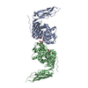
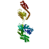
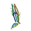
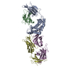
 Structure visualization
 Downloads & links
 Other downloads
 PDBj
PDBj


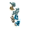
 Assembly
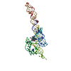

 Mass: 24.305 Da / Num. of mol.: 12 / Source method: obtained synthetically / Formula: Mg
 Mass: 161.116 Da / Num. of mol.: 7 / Source method: obtained synthetically / Formula: CoH18N6
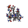
 Mass: 1580.590 Da / Num. of mol.: 1 / Source method: obtained synthetically / Formula: C72H101CoN18O17P / Comment: medication*YM
 Mass: 18.015 Da / Num. of mol.: 48 / Source method: isolated from a natural source / Formula: H2O
 Sample preparation
 Processing