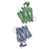
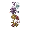
stress response to copper ion / sigma factor antagonist activity / protein unfolding / cellular response to unfolded protein / heat shock protein binding / protein folding chaperone / inclusion body / ATP-dependent protein folding chaperone / ADP binding / : ...stress response to copper ion / sigma factor antagonist activity / protein unfolding / cellular response to unfolded protein / heat shock protein binding / protein folding chaperone / inclusion body / ATP-dependent protein folding chaperone / ADP binding / : / response to heat / protein refolding / protein-folding chaperone binding / protein folding / protein-containing complex assembly / DNA replication / ATP hydrolysis activity / protein-containing complex / zinc ion binding / ATP binding / membrane / plasma membrane / cytoplasm / cytosol Similarity search - Function
Substrate Binding Domain Of Dnak; Chain:A; Domain 2 - #10 / Chaperone DnaK / Substrate Binding Domain Of DNAk; Chain A, domain 1 / Defensin A-like - #30 / Substrate Binding Domain Of DNAk; Chain A, domain 1 / Defensin A-like / Heat shock hsp70 proteins family signature 2. / Heat shock hsp70 proteins family signature 1. / Heat shock hsp70 proteins family signature 3. / Substrate Binding Domain Of Dnak; Chain:A; Domain 2 ...Substrate Binding Domain Of Dnak; Chain:A; Domain 2 - #10 / Chaperone DnaK / Substrate Binding Domain Of DNAk; Chain A, domain 1 / Defensin A-like - #30 / Substrate Binding Domain Of DNAk; Chain A, domain 1 / Defensin A-like / Heat shock hsp70 proteins family signature 2. / Heat shock hsp70 proteins family signature 1. / Heat shock hsp70 proteins family signature 3. / Substrate Binding Domain Of Dnak; Chain:A; Domain 2 / Heat shock protein 70, conserved site / Heat shock protein 70kD, peptide-binding domain superfamily / Heat shock protein 70kD, C-terminal domain superfamily / Heat shock protein 70 family / Hsp70 protein / ATPase, substrate binding domain, subdomain 4 / Actin; Chain A, domain 4 / ATPase, nucleotide binding domain / ATPase, nucleotide binding domain / Nucleotidyltransferase; domain 5 / Alpha-Beta Complex / Up-down Bundle / Sandwich / 2-Layer Sandwich / Mainly Beta / Mainly Alpha / Alpha Beta Similarity search - Domain/homology
Heatshockprotein70 / Chaperone protein dnaK / Heat shock 70 kDa protein / HSP70
Mass: 65651.023 Da / Num. of mol.: 1 Source method: isolated from a genetically manipulated source Source: (gene. exp.) Escherichia coli (E. coli) / Gene: dnaK, groP, grpF, seg, b0014, JW0013 / Production host: Escherichia coli (E. coli) / Strain (production host): BL2 / References: UniProt: P0A6Y8
Sequence details
THE SEQUENCE CONFLICT IN 1DKX, ASP530 WHICH SHOULD BE GLU530, HAS BEEN RETAINED IN THIS ENTRY.
-
Experimental details
-
Experiment
Experiment
Method: SOLUTION NMR
NMR experiment
Type: KAPPA-SHIFTED 15N-1H HSQC- TROSY
NMR details
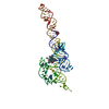
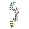
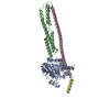
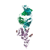
Text: NMR 1H-15N RESIDUAL DIPOLAR COUPLING (RDC) DATA IN 4% STRETCHED POLY ACRYL AMIDE. NH RDC DATA WERE OBTAINED FROM KAPPA-SHIFTED TROSY USING A VARIAN INOVA 800 MHZ NMR SPECTROMETER EQUIPPED WITH ...Text: NMR 1H-15N RESIDUAL DIPOLAR COUPLING (RDC) DATA IN 4% STRETCHED POLY ACRYL AMIDE. NH RDC DATA WERE OBTAINED FROM KAPPA-SHIFTED TROSY USING A VARIAN INOVA 800 MHZ NMR SPECTROMETER EQUIPPED WITH A HCN COLD (CRYO) PROBE. THE NUCLEOTIDE-BINDING DOMAIN (NBD) AND SUBSTRATE BINDING DOMAIN (SBD) ARE CONNECTED BY A FLEXIBLE LINKER AND MOVE WITH RESPECT TO EACHOTHER IN A CONE WITH AN ESTIMATED OPENING ANGLE OF 70 DEGREES ON THE NANO SECOND TIME SCALE.
-
Sample preparation
Details
Type: solution Contents: 0.2 MM [U-100% 13C U-100% 15N U-80% 2H] HSP70, 10 MM POTASSIUM CHLORIDE, 25 MM TRIS, 10 MM DTT, 5 MM MGCL2, 5 MM ADP, 10 MM POTASSIUM PHOSPHATE, 0.2 MM SODIUM AZIDE, 2 MM NRLLLTG, 90% H2O/ 10% D2O Label: sample_1 / Solvent system: 90% H2O/10% D2O
Method: RDC OPTIMIZATION / Software ordinal: 1 Details: RDC's WERE FITTED FOR (IA, IB, IIA) (IIB) (BETA, LID) AS SEPARATE UNITS USING GRID SEARCH OVER DA, DR, AND THREE EULER ANGLES FOLLOWED BY STEEPEST DESCEND. XRAY DATA FROM PDB ENTRY 1DKG WERE ...Details: RDC's WERE FITTED FOR (IA, IB, IIA) (IIB) (BETA, LID) AS SEPARATE UNITS USING GRID SEARCH OVER DA, DR, AND THREE EULER ANGLES FOLLOWED BY STEEPEST DESCEND. XRAY DATA FROM PDB ENTRY 1DKG WERE USED FOR RESIDUES 3-378 (NBD - NUCLEOTIDE BINDING DOMAIN). XRAY DATA FROM PDB ENTRY 1DKX WERE USED FOR 397-603 (SBD - SUBSTRATE BINDING DOMAIN). MISSING LOOPS IN 1DKG WERE ANNEALED AND MINIMIZED USING SWISSPROT SERVER. ORIENTATION OF DOMAIN IIB (RESIDUES 229-307) ADAPTED TO NMR DATA. DOMAIN IIB WAS ROTATED 20 DEGREES BASED ON RDC DATA AND MINIMALLY SUPERPOSED ON IIB IN 1DKG USING TRANSLATION AND ROTATION AROUND SZZ ONLY. CONNECTING RESIDUES 225-233 AND 307-314 WERE MINIMIZED IN SWISSPROT. NBD AND SBD WERE ORIENTED WITH RESPECT TO EACHOTHER BASED ON THE NMR DIPOLAR INFORMATION TRANSLATIONAL POSITION OF SBD WITH RESPECT TO NBD WAS DETERMINED BY COMPUTING THE BEST THEORETICAL ALLIGNMENT TENSOR USING PALES FROM ZWECKSTETTER M & BAX A (2001) J BIOMOL NMR 20(4):365-377. HETATM RECORDS IDENTIFY TENSOR ORIENTAIONS FOR DOMAINS IA,IB AND IIA AND BETA-LID OBTAINED FROM SELF_VALIDATION USING 50% OF THE RDC DATA. 65 A Z SEPARATION BETWEEN COORDINATE CENTERS OF NBD AND BETA-LID. 10 A Y SEPARATION BETWEEN COORDINATE CENTERS OF NBD AND BETA-LID. LINKER RESIDUES 379-395 ARE DYNAMIC AND WERE PLACED IN ARBITRARY CONFORMATION USING MOE (MOLECULAR OPERATING ENVIRONMENT) CHEMICAL COMPUTING GROUP 1010 SHERBROOKE ST. W, SUITE 910 MONTREAL, QUEBEC, CANADA H3A 2R7. LINKER CONFORMATION WAS OPTIMIZED IN MOE AND SWISS PROT.
NMR representative
Selection criteria: fewest violations
NMR ensemble
Conformer selection criteria: target function / Conformers calculated total number: 1 / Conformers submitted total number: 1
+
About Yorodumi
-
News
-
Feb 9, 2022. New format data for meta-information of EMDB entries
New format data for meta-information of EMDB entries
Version 3 of the EMDB header file is now the official format.
The previous official version 1.9 will be removed from the archive.
In the structure databanks used in Yorodumi, some data are registered as the other names, "COVID-19 virus" and "2019-nCoV". Here are the details of the virus and the list of structure data.
Jan 31, 2019. EMDB accession codes are about to change! (news from PDBe EMDB page)
EMDB accession codes are about to change! (news from PDBe EMDB page)
The allocation of 4 digits for EMDB accession codes will soon come to an end. Whilst these codes will remain in use, new EMDB accession codes will include an additional digit and will expand incrementally as the available range of codes is exhausted. The current 4-digit format prefixed with “EMD-” (i.e. EMD-XXXX) will advance to a 5-digit format (i.e. EMD-XXXXX), and so on. It is currently estimated that the 4-digit codes will be depleted around Spring 2019, at which point the 5-digit format will come into force.
The EM Navigator/Yorodumi systems omit the EMD- prefix.
Related info.:Q: What is EMD? / ID/Accession-code notation in Yorodumi/EM Navigator
Yorodumi is a browser for structure data from EMDB, PDB, SASBDB, etc.
This page is also the successor to EM Navigator detail page, and also detail information page/front-end page for Omokage search.
The word "yorodu" (or yorozu) is an old Japanese word meaning "ten thousand". "mi" (miru) is to see.
Related info.:EMDB / PDB / SASBDB / Comparison of 3 databanks / Yorodumi Search / Aug 31, 2016. New EM Navigator & Yorodumi / Yorodumi Papers / Jmol/JSmol / Function and homology information / Changes in new EM Navigator and Yorodumi
 Movie
Movie Controller

 Yorodumi
 Open data
 Basic information
 Components
 Keywords
 Function and homology information
 Authors
 Citation
 Structure visualization
 Downloads & links
 Other downloads
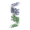
 PDBj
PDBj
 Assembly
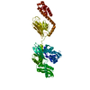
 HSQC- TROSY
 Sample preparation
 Processing