ムービー
ムービー コントローラー


+ データを開く

- 基本情報
基本情報
| 登録情報 | データベース: PDB / ID: 6vms | ||||||
|---|---|---|---|---|---|---|---|
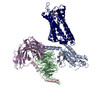
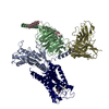
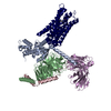
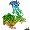
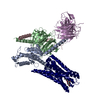
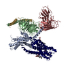
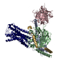
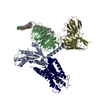
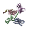
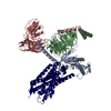
| タイトル | Structure of a D2 dopamine receptor-G-protein complex in a lipid membrane | ||||||
 要素 要素 |
| ||||||
 キーワード キーワード | SIGNALING PROTEIN / Dopamine / Dopamine receptor / GPCR / G protein / Parkinson's disease | ||||||
| 機能・相同性 |  機能・相同性情報 機能・相同性情報negative regulation of dopamine receptor signaling pathway / positive regulation of dopamine uptake involved in synaptic transmission / negative regulation of dephosphorylation / positive regulation of glial cell-derived neurotrophic factor production / acid secretion / dopamine neurotransmitter receptor activity, coupled via Gi/Go / nervous system process involved in regulation of systemic arterial blood pressure / response to histamine / negative regulation of circadian sleep/wake cycle, sleep / regulation of synapse structural plasticity ...negative regulation of dopamine receptor signaling pathway / positive regulation of dopamine uptake involved in synaptic transmission / negative regulation of dephosphorylation / positive regulation of glial cell-derived neurotrophic factor production / acid secretion / dopamine neurotransmitter receptor activity, coupled via Gi/Go / nervous system process involved in regulation of systemic arterial blood pressure / response to histamine / negative regulation of circadian sleep/wake cycle, sleep / regulation of synapse structural plasticity / regulation of locomotion involved in locomotory behavior / neuron-neuron synaptic transmission / adenylate cyclase regulator activity / Extra-nuclear estrogen signaling / Adenylate cyclase inhibitory pathway / negative regulation of cellular response to hypoxia / adenylate cyclase-inhibiting dopamine receptor signaling pathway / response to inactivity / regulation of potassium ion transport / orbitofrontal cortex development / negative regulation of dopamine secretion / adenohypophysis development / cerebral cortex GABAergic interneuron migration / negative regulation of neuron migration / hyaloid vascular plexus regression / Dopamine receptors / branching morphogenesis of a nerve / regulation of dopamine uptake involved in synaptic transmission / dopamine binding / positive regulation of growth hormone secretion / phospholipase C-activating dopamine receptor signaling pathway / heterotrimeric G-protein binding / peristalsis / beta-arrestin-dependent dopamine receptor signaling pathway / G protein-coupled receptor complex / grooming behavior / positive regulation of renal sodium excretion / drinking behavior / auditory behavior / striatum development / positive regulation of G protein-coupled receptor signaling pathway / dopaminergic synapse / behavioral response to ethanol / positive regulation of multicellular organism growth / G protein-coupled receptor internalization / non-motile cilium / negative regulation of synaptic transmission / Adrenaline,noradrenaline inhibits insulin secretion / ADP signalling through P2Y purinoceptor 12 / response to iron ion / positive regulation of urine volume / G alpha (i) signalling events / GTPase activating protein binding / adult walking behavior / negative regulation of synaptic transmission, glutamatergic / arachidonate secretion / ciliary membrane / positive regulation of neuroblast proliferation / response to morphine / regulation of synaptic transmission, GABAergic / neurotransmitter receptor localization to postsynaptic specialization membrane / positive regulation of cytokinesis / negative regulation of cytosolic calcium ion concentration / temperature homeostasis / pigmentation / dopamine metabolic process / dopamine uptake involved in synaptic transmission / regulation of dopamine secretion / positive regulation of receptor internalization / neuroblast proliferation / associative learning / lateral plasma membrane / cellular response to ethanol / negative regulation of protein secretion / response to light stimulus / G-protein alpha-subunit binding / potassium channel regulator activity / endocytic vesicle / prepulse inhibition / response to axon injury / sperm flagellum / long-term memory / viral release from host cell by cytolysis / postsynaptic modulation of chemical synaptic transmission / regulation of sodium ion transport / cellular response to retinoic acid / adenylate cyclase inhibitor activity / positive regulation of protein localization to cell cortex / negative regulation of blood pressure / T cell migration / peptidoglycan catabolic process / behavioral response to cocaine / release of sequestered calcium ion into cytosol / D2 dopamine receptor binding / synapse assembly / adenylate cyclase-inhibiting serotonin receptor signaling pathway / axonogenesis / G protein-coupled serotonin receptor binding / ionotropic glutamate receptor binding / epithelial cell proliferation 類似検索 - 分子機能 | ||||||
| 生物種 |   Homo sapiens (ヒト) Homo sapiens (ヒト) Enterobacteria phage T4 (ファージ) Enterobacteria phage T4 (ファージ) | ||||||
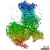
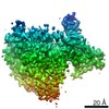
| 手法 | 電子顕微鏡法 / 単粒子再構成法 / クライオ電子顕微鏡法 / 解像度: 3.8 Å | ||||||
 データ登録者 データ登録者 | Yin, J. / Chen, K.M. / Clark, M.J. / Hijazi, M. / Kumari, P. / Bai, X. / Sunahara, R.K. / Barth, P. / Rosenbaum, D.M. | ||||||
| 資金援助 |  米国, 1件 米国, 1件
| ||||||
 引用 引用 | ジャーナル: Nature / 年: 2020 タイトル: Structure of a D2 dopamine receptor-G-protein complex in a lipid membrane. 著者: Jie Yin / Kuang-Yui M Chen / Mary J Clark / Mahdi Hijazi / Punita Kumari / Xiao-Chen Bai / Roger K Sunahara / Patrick Barth / Daniel M Rosenbaum /  要旨: The D2 dopamine receptor (DRD2) is a therapeutic target for Parkinson's disease and antipsychotic drugs. DRD2 is activated by the endogenous neurotransmitter dopamine and synthetic agonist drugs such ...The D2 dopamine receptor (DRD2) is a therapeutic target for Parkinson's disease and antipsychotic drugs. DRD2 is activated by the endogenous neurotransmitter dopamine and synthetic agonist drugs such as bromocriptine, leading to stimulation of G and inhibition of adenylyl cyclase. Here we used cryo-electron microscopy to elucidate the structure of an agonist-bound activated DRD2-G complex reconstituted into a phospholipid membrane. The extracellular ligand-binding site of DRD2 is remodelled in response to agonist binding, with conformational changes in extracellular loop 2, transmembrane domain 5 (TM5), TM6 and TM7, propagating to opening of the intracellular G-binding site. The DRD2-G structure represents, to our knowledge, the first experimental model of a G-protein-coupled receptor-G-protein complex embedded in a phospholipid bilayer, which serves as a benchmark to validate the interactions seen in previous detergent-bound structures. The structure also reveals interactions that are unique to the membrane-embedded complex, including helix 8 burial in the inner leaflet, ordered lysine and arginine side chains in the membrane interfacial regions, and lipid anchoring of the G protein in the membrane. Our model of the activated DRD2 will help to inform the design of subtype-selective DRD2 ligands for multiple human central nervous system disorders. | ||||||
| 履歴 |
|
- 構造の表示
構造の表示
| ムービー |
ムービービューア |
|---|---|
| 構造ビューア | 分子: MolmilJmol/JSmol |
- ダウンロードとリンク
-ダウンロード
| PDBx/mmCIF形式 | 6vms.cif.gz | 219.7 KB | 表示 | PDBx/mmCIF形式 |
|---|---|---|---|---|
| PDB形式 | pdb6vms.ent.gz | 167.5 KB | 表示 | PDB形式 |
| PDBx/mmJSON形式 | 6vms.json.gz | ツリー表示 | PDBx/mmJSON形式 | |
| その他 |  その他のダウンロード その他のダウンロード |
-検証レポート
| アーカイブディレクトリ | https://data.pdbj.org/pub/pdb/validation_reports/vm/6vmsftp://data.pdbj.org/pub/pdb/validation_reports/vm/6vms | HTTPS FTP |
|---|
-関連構造データ


-リンク
 PDBj
PDBj


- 集合体
集合体
| 登録構造単位 | 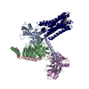
|
|---|---|
| 1 |
|
-要素
-Guanine nucleotide-binding protein ... , 3種, 3分子 ABC
| #1: タンパク質 | 分子量: 40382.047 Da / 分子数: 1 / 由来タイプ: 組換発現 / 由来: (組換発現) 発現宿主:   Spodoptera frugiperda (ツマジロクサヨトウ) Spodoptera frugiperda (ツマジロクサヨトウ)参照: UniProt: P10824 |
|---|---|
| #2: タンパク質 | 分子量: 37416.930 Da / 分子数: 1 / 由来タイプ: 組換発現 / 由来: (組換発現) Homo sapiens (ヒト) / 遺伝子: GNB1発現宿主: Spodoptera frugiperda (ツマジロクサヨトウ)参照: UniProt: P62873 |
| #3: タンパク質 | 分子量: 9137.474 Da / 分子数: 1 / 由来タイプ: 組換発現 / 由来: (組換発現) Homo sapiens (ヒト) / 遺伝子: GNG2発現宿主: Spodoptera frugiperda (ツマジロクサヨトウ)参照: UniProt: P59768 |
-抗体 / タンパク質 / 非ポリマー , 3種, 3分子 ER
| #4: 抗体 | 分子量: 27784.896 Da / 分子数: 1 / 由来タイプ: 組換発現 / 由来: (組換発現) Homo sapiens (ヒト)発現宿主: Spodoptera frugiperda (ツマジロクサヨトウ) |
|---|---|
| #5: タンパク質 | 分子量: 51384.059 Da / 分子数: 1 / 由来タイプ: 組換発現 由来: (組換発現) Enterobacteria phage T4 (ファージ), (組換発現) Homo sapiens (ヒト)遺伝子: e, T4Tp126, DRD2 発現宿主: Spodoptera frugiperda (ツマジロクサヨトウ)参照: UniProt: D9IEF7, UniProt: P14416, lysozyme |
| #6: 化合物 | ChemComp-08Y /  分子量: 654.594 Da / 分子数: 1 / 由来タイプ: 合成 / 式: C32H40BrN5O5 / タイプ: SUBJECT OF INVESTIGATION / コメント: アゴニスト*YM 分子量: 654.594 Da / 分子数: 1 / 由来タイプ: 合成 / 式: C32H40BrN5O5 / タイプ: SUBJECT OF INVESTIGATION / コメント: アゴニスト*YM |
-詳細
| 研究の焦点であるリガンドがあるか | Y |
|---|---|
| Has protein modification | Y |
-実験情報
-実験
| 実験 | 手法: 電子顕微鏡法 |
|---|---|
| EM実験 | 試料の集合状態: PARTICLE / 3次元再構成法: 単粒子再構成法 |
- 試料調製
試料調製
| 構成要素 | 名称: D2 dopamine receptor-G protein complex / タイプ: COMPLEX / Entity ID: #1-#5 / 由来: RECOMBINANT |
|---|---|
| 由来(天然) | 生物種: Homo sapiens (ヒト) |
| 由来(組換発現) | 生物種: Spodoptera frugiperda (ツマジロクサヨトウ) |
| 緩衝液 | pH: 7.4 |
| 試料 | 包埋: NO / シャドウイング: NO / 染色: NO / 凍結: YES |
| 急速凍結 | 凍結剤: ETHANE |
- 電子顕微鏡撮影
| 実験機器 |  モデル: Titan Krios / 画像提供: FEI Company |
|---|---|
| 顕微鏡 | モデル: FEI TITAN KRIOS |
| 電子銃 | 電子線源:  FIELD EMISSION GUN / 加速電圧: 300 kV / 照射モード: FLOOD BEAM FIELD EMISSION GUN / 加速電圧: 300 kV / 照射モード: FLOOD BEAM |
| 電子レンズ | モード: BRIGHT FIELD |
| 撮影 | 電子線照射量: 64 e/Å2 フィルム・検出器のモデル: GATAN K3 BIOQUANTUM (6k x 4k) |
- 解析
解析
| EMソフトウェア |
| ||||||||||||||||||||||||||||||||
|---|---|---|---|---|---|---|---|---|---|---|---|---|---|---|---|---|---|---|---|---|---|---|---|---|---|---|---|---|---|---|---|---|---|
| CTF補正 | タイプ: PHASE FLIPPING AND AMPLITUDE CORRECTION | ||||||||||||||||||||||||||||||||
| 対称性 | 点対称性: C1 (非対称) | ||||||||||||||||||||||||||||||||
| 3次元再構成 | 解像度: 3.8 Å / 解像度の算出法: FSC 0.143 CUT-OFF / 粒子像の数: 783984 / 対称性のタイプ: POINT | ||||||||||||||||||||||||||||||||
| 原子モデル構築 | PDB-ID: 6DDE Accession code: 6DDE / Source name: PDB / タイプ: experimental model |