[English] 日本語
 Yorodumi
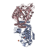
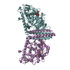
- PDB-6vlf: Crystal structure of mouse alpha 1,6-fucosyltransferase, FUT8 in ... -
+ Open data
- Basic information
| Entry | Database: PDB / ID: 6vlf | ||||||
|---|---|---|---|---|---|---|---|
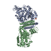
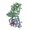
| Title | Crystal structure of mouse alpha 1,6-fucosyltransferase, FUT8 in its Apo-form | ||||||
 Components Components | Alpha-(1,6)-fucosyltransferase | ||||||
 Keywords Keywords | TRANSFERASE / FUT8 / Fucosyltransferase / Glycosyltransferase / N-glycan / Core fucose / SH3 domain | ||||||
| Function / homology |  Function and homology information Function and homology informationReactions specific to the complex N-glycan synthesis pathway / glycoprotein 6-alpha-L-fucosyltransferase / glycoprotein 6-alpha-L-fucosyltransferase activity / receptor metabolic process / GDP-L-fucose metabolic process / alpha-(1->6)-fucosyltransferase activity / N-glycan processing / regulation of cellular response to oxidative stress / respiratory gaseous exchange by respiratory system / : ...Reactions specific to the complex N-glycan synthesis pathway / glycoprotein 6-alpha-L-fucosyltransferase / glycoprotein 6-alpha-L-fucosyltransferase activity / receptor metabolic process / GDP-L-fucose metabolic process / alpha-(1->6)-fucosyltransferase activity / N-glycan processing / regulation of cellular response to oxidative stress / respiratory gaseous exchange by respiratory system / : / protein N-linked glycosylation / Golgi cisterna membrane / glycosyltransferase activity / transforming growth factor beta receptor signaling pathway / integrin-mediated signaling pathway / SH3 domain binding / cell migration / regulation of gene expression Similarity search - Function | ||||||
| Biological species |  | ||||||
| Method |  X-RAY DIFFRACTION / X-RAY DIFFRACTION /  SYNCHROTRON / SYNCHROTRON /  MOLECULAR REPLACEMENT / MOLECULAR REPLACEMENT /  molecular replacement / Resolution: 1.8 Å molecular replacement / Resolution: 1.8 Å | ||||||
 Authors Authors | Jarva, M.A. / Dramicanin, M. / Lingford, J.P. / Mao, R. / John, A. / Goddard-Borger, E. | ||||||
| Funding support |  Australia, 1items Australia, 1items
| ||||||
 Citation Citation |  Journal: J.Biol.Chem. / Year: 2020 Journal: J.Biol.Chem. / Year: 2020Title: Structural basis of substrate recognition and catalysis by fucosyltransferase 8. Authors: Jarva, M.A. / Dramicanin, M. / Lingford, J.P. / Mao, R. / John, A. / Jarman, K.E. / Grinter, R. / Goddard-Borger, E.D. | ||||||
| History |
|
- Structure visualization
| Structure viewer | Molecule:  Molmil Molmil Jmol/JSmol Jmol/JSmol |
|---|
- Downloads & links
- Download
| PDBx/mmCIF format |  6vlf.cif.gz 6vlf.cif.gz | 394.1 KB | Display |  PDBx/mmCIF format PDBx/mmCIF format |
|---|---|---|---|---|
| PDB format |  pdb6vlf.ent.gz pdb6vlf.ent.gz | 317.4 KB | Display |  PDB format PDB format |
| PDBx/mmJSON format |  6vlf.json.gz 6vlf.json.gz | Tree view |  PDBx/mmJSON format PDBx/mmJSON format | |
| Others |  Other downloads Other downloads |
-Validation report
| Arichive directory |  https://data.pdbj.org/pub/pdb/validation_reports/vl/6vlf https://data.pdbj.org/pub/pdb/validation_reports/vl/6vlf ftp://data.pdbj.org/pub/pdb/validation_reports/vl/6vlf ftp://data.pdbj.org/pub/pdb/validation_reports/vl/6vlf | HTTPS FTP |
|---|
-Related structure data
| Related structure data |  6vldC  6vleC 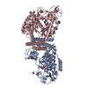 6vlgC  2de0S S: Starting model for refinement C: citing same article ( |
|---|---|
| Similar structure data |
- Links
Links
- Assembly
| Deposited unit | 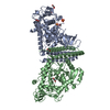
| ||||||||
|---|---|---|---|---|---|---|---|---|---|
| 1 |
| ||||||||
| Unit cell |
|
- Components
| #1: Protein | Mass: 62603.777 Da / Num. of mol.: 2 Source method: isolated from a genetically manipulated source Source: (gene. exp.)   References: UniProt: Q9WTS2, glycoprotein 6-alpha-L-fucosyltransferase #2: Chemical | ChemComp-SO4 / #3: Chemical | ChemComp-EDO / #4: Water | ChemComp-HOH / | Has ligand of interest | N | Has protein modification | Y | |
|---|
-Experimental details
-Experiment
| Experiment | Method:  X-RAY DIFFRACTION / Number of used crystals: 1 X-RAY DIFFRACTION / Number of used crystals: 1 |
|---|
- Sample preparation
| Crystal | Density Matthews: 2.7 Å3/Da / Density % sol: 54.48 % |
|---|---|
| Crystal grow | Temperature: 298 K / Method: vapor diffusion, sitting drop / pH: 7.4 / Details: 12% PEG 3350, 0.25 M NH4SO4, 0.1M Tris, pH 7.4 |
-Data collection
| Diffraction | Mean temperature: 100 K / Serial crystal experiment: N |
|---|---|
| Diffraction source | Source:  SYNCHROTRON / Site: SYNCHROTRON / Site:  Australian Synchrotron Australian Synchrotron  / Beamline: MX2 / Wavelength: 0.9537 Å / Beamline: MX2 / Wavelength: 0.9537 Å |
| Detector | Type: DECTRIS EIGER X 16M / Detector: PIXEL / Date: Aug 25, 2018 |
| Radiation | Protocol: SINGLE WAVELENGTH / Monochromatic (M) / Laue (L): M / Scattering type: x-ray |
| Radiation wavelength | Wavelength: 0.9537 Å / Relative weight: 1 |
| Reflection | Resolution: 1.8→49.2 Å / Num. obs: 123197 / % possible obs: 99.6 % / Redundancy: 3.4 % / CC1/2: 0.999 / Rmerge(I) obs: 0.052 / Rpim(I) all: 0.033 / Rrim(I) all: 0.062 / Net I/σ(I): 10.6 / Num. measured all: 422751 / Scaling rejects: 3 |
| Reflection shell | Resolution: 1.8→1.83 Å / Redundancy: 3.5 % / Rmerge(I) obs: 1.268 / Num. measured all: 21511 / Num. unique obs: 6122 / CC1/2: 0.463 / Rpim(I) all: 0.788 / Rrim(I) all: 1.496 / Net I/σ(I) obs: 0.9 / % possible all: 99.7 |
-Phasing
| Phasing | Method:  molecular replacement molecular replacement |
|---|
- Processing
| Software |
| ||||||||||||||||||||||||||||||||||||||||||||||||||||||||||||||||||||||||||||||||||||||||||||||||||||||||||||||||||||||||||||||||||||||||||||||||||||||||||||||||||||||||||||||||||||||||||
|---|---|---|---|---|---|---|---|---|---|---|---|---|---|---|---|---|---|---|---|---|---|---|---|---|---|---|---|---|---|---|---|---|---|---|---|---|---|---|---|---|---|---|---|---|---|---|---|---|---|---|---|---|---|---|---|---|---|---|---|---|---|---|---|---|---|---|---|---|---|---|---|---|---|---|---|---|---|---|---|---|---|---|---|---|---|---|---|---|---|---|---|---|---|---|---|---|---|---|---|---|---|---|---|---|---|---|---|---|---|---|---|---|---|---|---|---|---|---|---|---|---|---|---|---|---|---|---|---|---|---|---|---|---|---|---|---|---|---|---|---|---|---|---|---|---|---|---|---|---|---|---|---|---|---|---|---|---|---|---|---|---|---|---|---|---|---|---|---|---|---|---|---|---|---|---|---|---|---|---|---|---|---|---|---|---|---|---|
| Refinement | Method to determine structure:  MOLECULAR REPLACEMENT MOLECULAR REPLACEMENTStarting model: 2DE0 Resolution: 1.8→44.527 Å / SU ML: 0.23 / Cross valid method: THROUGHOUT / σ(F): 1.34 / Phase error: 23.72
| ||||||||||||||||||||||||||||||||||||||||||||||||||||||||||||||||||||||||||||||||||||||||||||||||||||||||||||||||||||||||||||||||||||||||||||||||||||||||||||||||||||||||||||||||||||||||||
| Solvent computation | Shrinkage radii: 0.9 Å / VDW probe radii: 1.11 Å | ||||||||||||||||||||||||||||||||||||||||||||||||||||||||||||||||||||||||||||||||||||||||||||||||||||||||||||||||||||||||||||||||||||||||||||||||||||||||||||||||||||||||||||||||||||||||||
| Displacement parameters | Biso max: 221.65 Å2 / Biso mean: 43.9381 Å2 / Biso min: 21.29 Å2 | ||||||||||||||||||||||||||||||||||||||||||||||||||||||||||||||||||||||||||||||||||||||||||||||||||||||||||||||||||||||||||||||||||||||||||||||||||||||||||||||||||||||||||||||||||||||||||
| Refinement step | Cycle: final / Resolution: 1.8→44.527 Å
| ||||||||||||||||||||||||||||||||||||||||||||||||||||||||||||||||||||||||||||||||||||||||||||||||||||||||||||||||||||||||||||||||||||||||||||||||||||||||||||||||||||||||||||||||||||||||||
| LS refinement shell | Refine-ID: X-RAY DIFFRACTION / Rfactor Rfree error: 0
| ||||||||||||||||||||||||||||||||||||||||||||||||||||||||||||||||||||||||||||||||||||||||||||||||||||||||||||||||||||||||||||||||||||||||||||||||||||||||||||||||||||||||||||||||||||||||||
| Refinement TLS params. | Method: refined / Refine-ID: X-RAY DIFFRACTION
| ||||||||||||||||||||||||||||||||||||||||||||||||||||||||||||||||||||||||||||||||||||||||||||||||||||||||||||||||||||||||||||||||||||||||||||||||||||||||||||||||||||||||||||||||||||||||||
| Refinement TLS group |
|
 Movie
Movie Controller




 PDBj
PDBj


