 Movie
Movie Controller

[English] 日本語
 Yorodumi
- PDB-6vkk: Crystal Structure of human PARP-1 CAT domain bound to inhibitor r... -
+ Open data

- Basic information
| Entry | Database: PDB / ID: 6vkk | ||||||
|---|---|---|---|---|---|---|---|
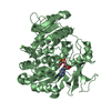
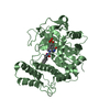
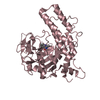
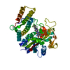
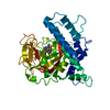
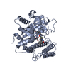
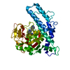
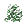
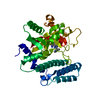
| Title | Crystal Structure of human PARP-1 CAT domain bound to inhibitor rucaparib | ||||||
 Components Components | Poly [ADP-ribose] polymerase 1 | ||||||
 Keywords Keywords | TRANSFERASE/INHIBITOR / PARP-1 / poly(ADP-ribose) polymerase / PARP inhibitor / PARP1 / ARTD1 / TRANSFERASE / TRANSFERASE-INHIBITOR complex | ||||||
| Function / homology |  Function and homology information Function and homology informationNAD+-histone H2BS6 serine ADP-ribosyltransferase activity / NAD+-histone H3S10 serine ADP-ribosyltransferase activity / NAD+-histone H2BE35 glutamate ADP-ribosyltransferase activity / positive regulation of myofibroblast differentiation / negative regulation of ATP biosynthetic process / NAD+-protein-tyrosine ADP-ribosyltransferase activity / NAD+-protein-histidine ADP-ribosyltransferase activity / regulation of base-excision repair / regulation of circadian sleep/wake cycle, non-REM sleep / mitochondrial DNA metabolic process ...NAD+-histone H2BS6 serine ADP-ribosyltransferase activity / NAD+-histone H3S10 serine ADP-ribosyltransferase activity / NAD+-histone H2BE35 glutamate ADP-ribosyltransferase activity / positive regulation of myofibroblast differentiation / negative regulation of ATP biosynthetic process / NAD+-protein-tyrosine ADP-ribosyltransferase activity / NAD+-protein-histidine ADP-ribosyltransferase activity / regulation of base-excision repair / regulation of circadian sleep/wake cycle, non-REM sleep / mitochondrial DNA metabolic process / vRNA Synthesis / carbohydrate biosynthetic process / NAD+-protein-serine ADP-ribosyltransferase activity / NAD DNA ADP-ribosyltransferase activity / negative regulation of adipose tissue development / DNA ADP-ribosylation / regulation of oxidative stress-induced neuron intrinsic apoptotic signaling pathway / ATP generation from poly-ADP-D-ribose / replication fork reversal / positive regulation of necroptotic process / signal transduction involved in regulation of gene expression / transcription regulator activator activity / response to aldosterone / HDR through MMEJ (alt-NHEJ) / positive regulation of DNA-templated transcription, elongation / NAD+ ADP-ribosyltransferase / protein auto-ADP-ribosylation / negative regulation of telomere maintenance via telomere lengthening / NAD+-protein-aspartate ADP-ribosyltransferase activity / mitochondrial DNA repair / positive regulation of intracellular estrogen receptor signaling pathway / protein poly-ADP-ribosylation / NAD+-protein-glutamate ADP-ribosyltransferase activity / negative regulation of cGAS/STING signaling pathway / positive regulation of cardiac muscle hypertrophy / NAD+-protein mono-ADP-ribosyltransferase activity / decidualization / cellular response to zinc ion / protein autoprocessing / positive regulation of mitochondrial depolarization / nuclear replication fork / R-SMAD binding / Transferases; Glycosyltransferases; Pentosyltransferases / positive regulation of SMAD protein signal transduction / negative regulation of transcription elongation by RNA polymerase II / macrophage differentiation / POLB-Dependent Long Patch Base Excision Repair / NAD+ poly-ADP-ribosyltransferase activity / SUMOylation of DNA damage response and repair proteins / nucleosome binding / positive regulation of double-strand break repair via homologous recombination / site of DNA damage / protein localization to chromatin / nucleotidyltransferase activity / positive regulation of adipose tissue development / transforming growth factor beta receptor signaling pathway / negative regulation of innate immune response / telomere maintenance / nuclear estrogen receptor binding / protein modification process / response to gamma radiation / mitochondrion organization / Downregulation of SMAD2/3:SMAD4 transcriptional activity / cellular response to nerve growth factor stimulus / protein-DNA complex / positive regulation of protein localization to nucleus / DNA Damage Recognition in GG-NER / NAD binding / cellular response to amyloid-beta / enzyme activator activity / histone deacetylase binding / Dual Incision in GG-NER / cellular response to insulin stimulus / Formation of Incision Complex in GG-NER / cellular response to UV / nuclear envelope / double-strand break repair / regulation of protein localization / site of double-strand break / cellular response to oxidative stress / transcription regulator complex / transcription by RNA polymerase II / damaged DNA binding / RNA polymerase II-specific DNA-binding transcription factor binding / response to ethanol / positive regulation of canonical NF-kappaB signal transduction / chromosome, telomeric region / nuclear body / innate immune response / DNA repair / negative regulation of DNA-templated transcription / apoptotic process / chromatin binding / DNA damage response / ubiquitin protein ligase binding / protein kinase binding / chromatin / nucleolus / enzyme binding / negative regulation of transcription by RNA polymerase II Similarity search - Function | ||||||
| Biological species |  Homo sapiens (human) Homo sapiens (human) | ||||||
| Method |  X-RAY DIFFRACTION / SYNCHROTRON / MOLECULAR REPLACEMENT / molecular replacement / Resolution: 2.1 Å X-RAY DIFFRACTION / SYNCHROTRON / MOLECULAR REPLACEMENT / molecular replacement / Resolution: 2.1 Å | ||||||
 Authors Authors | Steffen, J.D. / Pascal, J.M. | ||||||
| Funding support |  Canada, 1items Canada, 1items
| ||||||
 Citation Citation | Journal: Science / Year: 2020 Title: Structural basis for allosteric PARP-1 retention on DNA breaks. Authors: Zandarashvili, L. / Langelier, M.F. / Velagapudi, U.K. / Hancock, M.A. / Steffen, J.D. / Billur, R. / Hannan, Z.M. / Wicks, A.J. / Krastev, D.B. / Pettitt, S.J. / Lord, C.J. / Talele, T.T. / ...Authors: Zandarashvili, L. / Langelier, M.F. / Velagapudi, U.K. / Hancock, M.A. / Steffen, J.D. / Billur, R. / Hannan, Z.M. / Wicks, A.J. / Krastev, D.B. / Pettitt, S.J. / Lord, C.J. / Talele, T.T. / Pascal, J.M. / Black, B.E. | ||||||
| History |
|
- Structure visualization
| Structure viewer | Molecule: MolmilJmol/JSmol |
|---|
- Downloads & links
-Download
| PDBx/mmCIF format | 6vkk.cif.gz | 550.3 KB | Display | PDBx/mmCIF format |
|---|---|---|---|---|
| PDB format | pdb6vkk.ent.gz | 455.3 KB | Display | PDB format |
| PDBx/mmJSON format | 6vkk.json.gz | Tree view | PDBx/mmJSON format | |
| Others |  Other downloads Other downloads |
-Validation report
| Arichive directory | https://data.pdbj.org/pub/pdb/validation_reports/vk/6vkkftp://data.pdbj.org/pub/pdb/validation_reports/vk/6vkk | HTTPS FTP |
|---|
-Related structure data
| Related structure data |  6ntuC  6vkoC 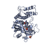 6vkqC 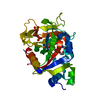 5ds3S S: Starting model for refinement C: citing same article ( |
|---|---|
| Similar structure data |

-Links
 PDBj
PDBj


- Assembly
| Deposited unit | 
| ||||||||||||||||||||||||||||||||||||||||||||||||||||||||||||||||||||||||||||||||||||||||||||||||||
|---|---|---|---|---|---|---|---|---|---|---|---|---|---|---|---|---|---|---|---|---|---|---|---|---|---|---|---|---|---|---|---|---|---|---|---|---|---|---|---|---|---|---|---|---|---|---|---|---|---|---|---|---|---|---|---|---|---|---|---|---|---|---|---|---|---|---|---|---|---|---|---|---|---|---|---|---|---|---|---|---|---|---|---|---|---|---|---|---|---|---|---|---|---|---|---|---|---|---|---|
| 1 | 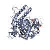
| ||||||||||||||||||||||||||||||||||||||||||||||||||||||||||||||||||||||||||||||||||||||||||||||||||
| 2 | 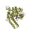
| ||||||||||||||||||||||||||||||||||||||||||||||||||||||||||||||||||||||||||||||||||||||||||||||||||
| 3 | 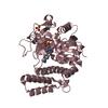
| ||||||||||||||||||||||||||||||||||||||||||||||||||||||||||||||||||||||||||||||||||||||||||||||||||
| 4 | 
| ||||||||||||||||||||||||||||||||||||||||||||||||||||||||||||||||||||||||||||||||||||||||||||||||||
| Unit cell |
| ||||||||||||||||||||||||||||||||||||||||||||||||||||||||||||||||||||||||||||||||||||||||||||||||||
| Noncrystallographic symmetry (NCS) | NCS domain:
NCS domain segments: Component-ID: _ / Beg auth comp-ID: LYS / Beg label comp-ID: LYS / End auth comp-ID: THR / End label comp-ID: THR / Refine code: _ / Auth seq-ID: 662 - 1011 / Label seq-ID: 23 - 372
NCS ensembles :
|
-Components
| #1: Protein | Mass: 41572.461 Da / Num. of mol.: 4 / Fragment: catalytic domain Source method: isolated from a genetically manipulated source Source: (gene. exp.) Homo sapiens (human) / Gene: PARP1, ADPRT, PPOL / Production host:  References: UniProt: P09874, NAD+ ADP-ribosyltransferase, Transferases; Glycosyltransferases; Pentosyltransferases #2: Chemical | ChemComp-RPB /   Mass: 323.364 Da / Num. of mol.: 4 / Source method: obtained synthetically / Formula: C19H18FN3O / Feature type: SUBJECT OF INVESTIGATION Mass: 323.364 Da / Num. of mol.: 4 / Source method: obtained synthetically / Formula: C19H18FN3O / Feature type: SUBJECT OF INVESTIGATION#3: Chemical |   Mass: 92.094 Da / Num. of mol.: 3 / Source method: obtained synthetically / Formula: C3H8O3 Mass: 92.094 Da / Num. of mol.: 3 / Source method: obtained synthetically / Formula: C3H8O3#4: Chemical | ChemComp-SO4 /   Mass: 96.063 Da / Num. of mol.: 9 / Source method: obtained synthetically / Formula: SO4 Mass: 96.063 Da / Num. of mol.: 9 / Source method: obtained synthetically / Formula: SO4#5: Water | ChemComp-HOH / |  Mass: 18.015 Da / Num. of mol.: 169 / Source method: isolated from a natural source / Formula: H2O Mass: 18.015 Da / Num. of mol.: 169 / Source method: isolated from a natural source / Formula: H2OHas ligand of interest | Y | Has protein modification | Y | |
|---|
-Experimental details
-Experiment
| Experiment | Method: X-RAY DIFFRACTION / Number of used crystals: 1 |
|---|
- Sample preparation
| Crystal | Density Matthews: 2.42 Å3/Da / Density % sol: 49.09 % |
|---|---|
| Crystal grow | Temperature: 298 K / Method: vapor diffusion, sitting drop / pH: 8 / Details: 2M Ammonium Sulfate, 5% PEG 400, 100 mM Tris |
-Data collection
| Diffraction | Mean temperature: 100 K / Serial crystal experiment: N | ||||||||||||||||||||||||||||||
|---|---|---|---|---|---|---|---|---|---|---|---|---|---|---|---|---|---|---|---|---|---|---|---|---|---|---|---|---|---|---|---|
| Diffraction source | Source: SYNCHROTRON / Site: ALS  / Beamline: 12.3.1 / Wavelength: 1.1158 Å / Beamline: 12.3.1 / Wavelength: 1.1158 Å | ||||||||||||||||||||||||||||||
| Detector | Type: ADSC QUANTUM 270 / Detector: CCD / Date: Aug 1, 2014 | ||||||||||||||||||||||||||||||
| Radiation | Protocol: SINGLE WAVELENGTH / Monochromatic (M) / Laue (L): M / Scattering type: x-ray | ||||||||||||||||||||||||||||||
| Radiation wavelength | Wavelength: 1.1158 Å / Relative weight: 1 | ||||||||||||||||||||||||||||||
| Reflection | Resolution: 2.1→47.92 Å / Num. obs: 94458 / % possible obs: 99.8 % / Redundancy: 11.7 % / CC1/2: 0.998 / Rmerge(I) obs: 0.089 / Rpim(I) all: 0.028 / Rrim(I) all: 0.094 / Net I/σ(I): 16.3 / Num. measured all: 1105071 / Scaling rejects: 81 | ||||||||||||||||||||||||||||||
| Reflection shell | Diffraction-ID: 1
|
-Phasing
| Phasing | Method: molecular replacement |
|---|
- Processing
| Software |
| |||||||||||||||||||||||||||||||||||||||||||||||||||||||||||||||||||||||||||||||||||||||||||||||||||||||||||||||||||||||||||||||||||||||||||||||||||||||||||||||||||||||||||||||||||||||||||||||||||||||||||||||||||||||||||||||||
|---|---|---|---|---|---|---|---|---|---|---|---|---|---|---|---|---|---|---|---|---|---|---|---|---|---|---|---|---|---|---|---|---|---|---|---|---|---|---|---|---|---|---|---|---|---|---|---|---|---|---|---|---|---|---|---|---|---|---|---|---|---|---|---|---|---|---|---|---|---|---|---|---|---|---|---|---|---|---|---|---|---|---|---|---|---|---|---|---|---|---|---|---|---|---|---|---|---|---|---|---|---|---|---|---|---|---|---|---|---|---|---|---|---|---|---|---|---|---|---|---|---|---|---|---|---|---|---|---|---|---|---|---|---|---|---|---|---|---|---|---|---|---|---|---|---|---|---|---|---|---|---|---|---|---|---|---|---|---|---|---|---|---|---|---|---|---|---|---|---|---|---|---|---|---|---|---|---|---|---|---|---|---|---|---|---|---|---|---|---|---|---|---|---|---|---|---|---|---|---|---|---|---|---|---|---|---|---|---|---|---|---|---|---|---|---|---|---|---|---|---|---|---|---|---|---|---|
| Refinement | Method to determine structure: MOLECULAR REPLACEMENT Starting model: 5ds3 Resolution: 2.1→20 Å / Cor.coef. Fo:Fc: 0.942 / Cor.coef. Fo:Fc free: 0.929 / SU B: 13.189 / SU ML: 0.169 / SU R Cruickshank DPI: 0.2562 / Cross valid method: THROUGHOUT / σ(F): 0 / ESU R: 0.256 / ESU R Free: 0.197 Details: HYDROGENS HAVE BEEN ADDED IN THE RIDING POSITIONS U VALUES : WITH TLS ADDED
| |||||||||||||||||||||||||||||||||||||||||||||||||||||||||||||||||||||||||||||||||||||||||||||||||||||||||||||||||||||||||||||||||||||||||||||||||||||||||||||||||||||||||||||||||||||||||||||||||||||||||||||||||||||||||||||||||
| Solvent computation | Ion probe radii: 0.8 Å / Shrinkage radii: 0.8 Å / VDW probe radii: 1.2 Å | |||||||||||||||||||||||||||||||||||||||||||||||||||||||||||||||||||||||||||||||||||||||||||||||||||||||||||||||||||||||||||||||||||||||||||||||||||||||||||||||||||||||||||||||||||||||||||||||||||||||||||||||||||||||||||||||||
| Displacement parameters | Biso max: 145.22 Å2 / Biso mean: 55.022 Å2 / Biso min: 29.15 Å2
| |||||||||||||||||||||||||||||||||||||||||||||||||||||||||||||||||||||||||||||||||||||||||||||||||||||||||||||||||||||||||||||||||||||||||||||||||||||||||||||||||||||||||||||||||||||||||||||||||||||||||||||||||||||||||||||||||
| Refinement step | Cycle: final / Resolution: 2.1→20 Å
| |||||||||||||||||||||||||||||||||||||||||||||||||||||||||||||||||||||||||||||||||||||||||||||||||||||||||||||||||||||||||||||||||||||||||||||||||||||||||||||||||||||||||||||||||||||||||||||||||||||||||||||||||||||||||||||||||
| Refine LS restraints |
| |||||||||||||||||||||||||||||||||||||||||||||||||||||||||||||||||||||||||||||||||||||||||||||||||||||||||||||||||||||||||||||||||||||||||||||||||||||||||||||||||||||||||||||||||||||||||||||||||||||||||||||||||||||||||||||||||
| Refine LS restraints NCS | Refine-ID: X-RAY DIFFRACTION / Type: interatomic distance / Weight position: 0.05
| |||||||||||||||||||||||||||||||||||||||||||||||||||||||||||||||||||||||||||||||||||||||||||||||||||||||||||||||||||||||||||||||||||||||||||||||||||||||||||||||||||||||||||||||||||||||||||||||||||||||||||||||||||||||||||||||||
| LS refinement shell | Resolution: 2.1→2.154 Å / Rfactor Rfree error: 0 / Total num. of bins used: 20
| |||||||||||||||||||||||||||||||||||||||||||||||||||||||||||||||||||||||||||||||||||||||||||||||||||||||||||||||||||||||||||||||||||||||||||||||||||||||||||||||||||||||||||||||||||||||||||||||||||||||||||||||||||||||||||||||||
| Refinement TLS params. | Method: refined / Refine-ID: X-RAY DIFFRACTION
| |||||||||||||||||||||||||||||||||||||||||||||||||||||||||||||||||||||||||||||||||||||||||||||||||||||||||||||||||||||||||||||||||||||||||||||||||||||||||||||||||||||||||||||||||||||||||||||||||||||||||||||||||||||||||||||||||
| Refinement TLS group |
|
