 Movie
Movie Controller

[English] 日本語
 Yorodumi
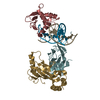
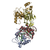
- PDB-6vge: Crystal structure of the DNA binding domains of human transcripti... -
+ Open data

- Basic information
| Entry | Database: PDB / ID: 6vge | |||||||||
|---|---|---|---|---|---|---|---|---|---|---|
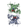
| Title | Crystal structure of the DNA binding domains of human transcription factor ERG, human Runx2 bound to core binding factor beta (Cbfb), in complex with 16mer DNA CAGAGGATGTGGCTTC | |||||||||
 Components Components |
| |||||||||
 Keywords Keywords | TRANSCRIPTION / Ewing sarcoma / enhancer / transcription factor / oncogenesis / prostate cancer / ETS-family / Runt-family | |||||||||
| Function / homology |  Function and homology information Function and homology informationligamentous ossification / regulation of fibroblast growth factor receptor signaling pathway / osteoblast fate commitment / RUNX3 regulates RUNX1-mediated transcription / RUNX1 regulates transcription of genes involved in BCR signaling / regulation of odontogenesis of dentin-containing tooth / chondrocyte development / RUNX1 regulates transcription of genes involved in interleukin signaling / RUNX2 regulates bone development / core-binding factor complex ...ligamentous ossification / regulation of fibroblast growth factor receptor signaling pathway / osteoblast fate commitment / RUNX3 regulates RUNX1-mediated transcription / RUNX1 regulates transcription of genes involved in BCR signaling / regulation of odontogenesis of dentin-containing tooth / chondrocyte development / RUNX1 regulates transcription of genes involved in interleukin signaling / RUNX2 regulates bone development / core-binding factor complex / RUNX1 regulates expression of components of tight junctions / positive regulation of CD8-positive, alpha-beta T cell differentiation / RUNX2 regulates chondrocyte maturation / negative regulation of CD4-positive, alpha-beta T cell differentiation / bHLH transcription factor binding / positive regulation of chondrocyte differentiation / RUNX1 and FOXP3 control the development of regulatory T lymphocytes (Tregs) / response to sodium phosphate / lymphocyte differentiation / RUNX2 regulates genes involved in cell migration / Transcriptional regulation by RUNX2 / YAP1- and WWTR1 (TAZ)-stimulated gene expression / RUNX2 regulates genes involved in differentiation of myeloid cells / RUNX1 regulates transcription of genes involved in differentiation of keratinocytes / endochondral ossification / SMAD protein signal transduction / embryonic cranial skeleton morphogenesis / RUNX3 Regulates Immune Response and Cell Migration / myeloid cell differentiation / definitive hemopoiesis / embryonic forelimb morphogenesis / RUNX1 regulates transcription of genes involved in differentiation of myeloid cells / Regulation of RUNX1 Expression and Activity / osteoblast development / RUNX1 regulates transcription of genes involved in WNT signaling / RUNX1 regulates estrogen receptor mediated transcription / smoothened signaling pathway / positive regulation of stem cell proliferation / RUNX2 regulates osteoblast differentiation / odontogenesis of dentin-containing tooth / bone mineralization / RUNX1 interacts with co-factors whose precise effect on RUNX1 targets is not known / regulation of cell differentiation / hemopoiesis / T cell differentiation / regulation of ossification / RUNX3 regulates p14-ARF / chondrocyte differentiation / positive regulation of osteoblast differentiation / BMP signaling pathway / cell maturation / epithelial cell proliferation / ossification / stem cell proliferation / positive regulation of epithelial cell proliferation / stem cell differentiation / negative regulation of smoothened signaling pathway / RUNX1 regulates genes involved in megakaryocyte differentiation and platelet function / chromatin DNA binding / Regulation of RUNX3 expression and activity / Transcriptional regulation of granulopoiesis / neuron differentiation / sequence-specific double-stranded DNA binding / protein polyubiquitination / osteoblast differentiation / Regulation of RUNX2 expression and activity / RUNX1 regulates transcription of genes involved in differentiation of HSCs / DNA-binding transcription activator activity, RNA polymerase II-specific / transcription regulator complex / transcription by RNA polymerase II / Estrogen-dependent gene expression / sequence-specific DNA binding / gene expression / protein phosphorylation / DNA-binding transcription factor activity, RNA polymerase II-specific / cell differentiation / transcription coactivator activity / RNA polymerase II cis-regulatory region sequence-specific DNA binding / DNA-binding transcription factor activity / ribonucleoprotein complex / protein domain specific binding / negative regulation of DNA-templated transcription / chromatin binding / positive regulation of gene expression / regulation of transcription by RNA polymerase II / positive regulation of DNA-templated transcription / chromatin / negative regulation of transcription by RNA polymerase II / signal transduction / positive regulation of transcription by RNA polymerase II / DNA binding / nucleoplasm / ATP binding / membrane / nucleus / cytoplasm / cytosol Similarity search - Function | |||||||||
| Biological species |  Homo sapiens (human) Homo sapiens (human) | |||||||||
| Method |  X-RAY DIFFRACTION / SYNCHROTRON / MOLECULAR REPLACEMENT / Resolution: 4.25 Å X-RAY DIFFRACTION / SYNCHROTRON / MOLECULAR REPLACEMENT / Resolution: 4.25 Å | |||||||||
 Authors Authors | Hou, C. / Tsodikov, O.V. | |||||||||
| Funding support |  United States, 2items United States, 2items
| |||||||||
 Citation Citation | Journal: Structure / Year: 2021 Title: Allosteric interference in oncogenic FLI1 and ERG transactions by mithramycins. Authors: Hou, C. / Mandal, A. / Rohr, J. / Tsodikov, O.V. | |||||||||
| History |
|
- Structure visualization
| Structure viewer | Molecule: MolmilJmol/JSmol |
|---|
- Downloads & links
-Download
| PDBx/mmCIF format | 6vge.cif.gz | 195.5 KB | Display | PDBx/mmCIF format |
|---|---|---|---|---|
| PDB format | pdb6vge.ent.gz | 152.8 KB | Display | PDB format |
| PDBx/mmJSON format | 6vge.json.gz | Tree view | PDBx/mmJSON format | |
| Others |  Other downloads Other downloads |
-Validation report
| Arichive directory | https://data.pdbj.org/pub/pdb/validation_reports/vg/6vgeftp://data.pdbj.org/pub/pdb/validation_reports/vg/6vge | HTTPS FTP |
|---|
-Related structure data
| Related structure data |  6vg2C 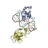 6vg8C 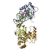 6vgdSC  6vggC S: Starting model for refinement C: citing same article ( |
|---|---|
| Similar structure data |


-Links
 PDBj
PDBj


- Assembly
| Deposited unit | 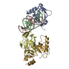
| ||||||||
|---|---|---|---|---|---|---|---|---|---|
| 1 |
| ||||||||
| Unit cell |
|
-Components
| #1: Protein | Mass: 14927.827 Da / Num. of mol.: 1 / Fragment: DNA binding domain Source method: isolated from a genetically manipulated source Details: The N-terminal GPHM is left over from the affinity tag after its cleavage Source: (gene. exp.) Homo sapiens (human) / Gene: ERG / Production host:  |
|---|---|
| #2: DNA chain | Mass: 4954.214 Da / Num. of mol.: 1 / Source method: obtained synthetically / Details: Synthetic DNA / Source: (synth.) Homo sapiens (human) |
| #3: DNA chain | Mass: 4843.156 Da / Num. of mol.: 1 / Source method: obtained synthetically / Details: Synthetic DNA / Source: (synth.) Homo sapiens (human) |
| #4: Protein | Mass: 19542.148 Da / Num. of mol.: 1 / Fragment: DNA binding domain Source method: isolated from a genetically manipulated source Source: (gene. exp.) Homo sapiens (human) / Gene: RUNX2, AML3, CBFA1, OSF2, PEBP2A / Production host: |
| #5: Protein | Mass: 18263.381 Da / Num. of mol.: 1 / Fragment: DNA binding domain Source method: isolated from a genetically manipulated source Details: The N-terminal AGSSHHHHHHSQDP is the affinity tag. The gaps correspond to disorder in the crystal. Source: (gene. exp.) Homo sapiens (human) / Gene: CBFB / Production host: |
-Experimental details
-Experiment
| Experiment | Method: X-RAY DIFFRACTION / Number of used crystals: 1 |
|---|
- Sample preparation
| Crystal | Density Matthews: 4.03 Å3/Da / Density % sol: 69.47 % |
|---|---|
| Crystal grow | Temperature: 294 K / Method: vapor diffusion, hanging drop / Details: 0.6 M K/Na Tartrate, 0.1 M Hepes, pH7.0 |
-Data collection
| Diffraction | Mean temperature: 100 K / Serial crystal experiment: N |
|---|---|
| Diffraction source | Source: SYNCHROTRON / Site: APS / Beamline: 22-ID / Wavelength: 1 Å |
| Detector | Type: RAYONIX MX300-HS / Detector: CCD / Date: Oct 22, 2016 |
| Radiation | Protocol: SINGLE WAVELENGTH / Monochromatic (M) / Laue (L): M / Scattering type: x-ray |
| Radiation wavelength | Wavelength: 1 Å / Relative weight: 1 |
| Reflection | Resolution: 4.25→50 Å / Num. obs: 7550 / % possible obs: 94.7 % / Redundancy: 6.1 % / Rmerge(I) obs: 0.103 / Net I/σ(I): 17.9 |
| Reflection shell | Resolution: 4.25→4.32 Å / Rmerge(I) obs: 1.382 / Num. unique obs: 367 / CC1/2: 0.785 / CC star: 0.938 |
- Processing
| Software |
| ||||||||||||||||||||||||||||||||||||||||||||||||||||||||||||||||||||||||||||||||||||||||||||||||||||||||||||||||||||||||||||||||||||||||||||||||||||||
|---|---|---|---|---|---|---|---|---|---|---|---|---|---|---|---|---|---|---|---|---|---|---|---|---|---|---|---|---|---|---|---|---|---|---|---|---|---|---|---|---|---|---|---|---|---|---|---|---|---|---|---|---|---|---|---|---|---|---|---|---|---|---|---|---|---|---|---|---|---|---|---|---|---|---|---|---|---|---|---|---|---|---|---|---|---|---|---|---|---|---|---|---|---|---|---|---|---|---|---|---|---|---|---|---|---|---|---|---|---|---|---|---|---|---|---|---|---|---|---|---|---|---|---|---|---|---|---|---|---|---|---|---|---|---|---|---|---|---|---|---|---|---|---|---|---|---|---|---|---|---|---|
| Refinement | Method to determine structure: MOLECULAR REPLACEMENT Starting model: 6VGD Resolution: 4.25→35 Å / Cor.coef. Fo:Fc: 0.924 / Cor.coef. Fo:Fc free: 0.907 / SU B: 240.05 / SU ML: 1.18 / Cross valid method: THROUGHOUT / σ(F): 0 / ESU R Free: 0.96 Details: HYDROGENS HAVE BEEN ADDED IN THE RIDING POSITIONS U VALUES : WITH TLS ADDED
| ||||||||||||||||||||||||||||||||||||||||||||||||||||||||||||||||||||||||||||||||||||||||||||||||||||||||||||||||||||||||||||||||||||||||||||||||||||||
| Solvent computation | Ion probe radii: 0.8 Å / Shrinkage radii: 0.8 Å / VDW probe radii: 1.2 Å | ||||||||||||||||||||||||||||||||||||||||||||||||||||||||||||||||||||||||||||||||||||||||||||||||||||||||||||||||||||||||||||||||||||||||||||||||||||||
| Displacement parameters | Biso max: 438.53 Å2 / Biso mean: 282.708 Å2 / Biso min: 189.17 Å2
| ||||||||||||||||||||||||||||||||||||||||||||||||||||||||||||||||||||||||||||||||||||||||||||||||||||||||||||||||||||||||||||||||||||||||||||||||||||||
| Refinement step | Cycle: final / Resolution: 4.25→35 Å
| ||||||||||||||||||||||||||||||||||||||||||||||||||||||||||||||||||||||||||||||||||||||||||||||||||||||||||||||||||||||||||||||||||||||||||||||||||||||
| Refine LS restraints |
| ||||||||||||||||||||||||||||||||||||||||||||||||||||||||||||||||||||||||||||||||||||||||||||||||||||||||||||||||||||||||||||||||||||||||||||||||||||||
| LS refinement shell | Resolution: 4.25→4.359 Å / Rfactor Rfree error: 0 / Total num. of bins used: 20
| ||||||||||||||||||||||||||||||||||||||||||||||||||||||||||||||||||||||||||||||||||||||||||||||||||||||||||||||||||||||||||||||||||||||||||||||||||||||
| Refinement TLS params. | Method: refined / Refine-ID: X-RAY DIFFRACTION
| ||||||||||||||||||||||||||||||||||||||||||||||||||||||||||||||||||||||||||||||||||||||||||||||||||||||||||||||||||||||||||||||||||||||||||||||||||||||
| Refinement TLS group |
|
