 Movie
Movie Controller


+ Open data

- Basic information
| Entry | Database: PDB / ID: 6vfv | ||||||||||||
|---|---|---|---|---|---|---|---|---|---|---|---|---|---|
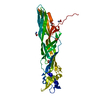
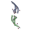
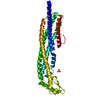
| Title | Crystal structure of human protocadherin 8 EC5-EC6 | ||||||||||||
 Components Components | Protocadherin-8 | ||||||||||||
 Keywords Keywords | CELL ADHESION / cadherin extracellular region / non-clustered delta2 protocadherin / homophilic adhesion/recognition calcium-dependent adhesion molecule | ||||||||||||
| Function / homology |  Function and homology information Function and homology informationmorphogenesis of embryonic epithelium / regulation of synaptic membrane adhesion / homophilic cell-cell adhesion / somitogenesis / modulation of chemical synaptic transmission / Schaffer collateral - CA1 synapse / cell-cell signaling / presynaptic membrane / chemical synaptic transmission / postsynaptic membrane ...morphogenesis of embryonic epithelium / regulation of synaptic membrane adhesion / homophilic cell-cell adhesion / somitogenesis / modulation of chemical synaptic transmission / Schaffer collateral - CA1 synapse / cell-cell signaling / presynaptic membrane / chemical synaptic transmission / postsynaptic membrane / cell adhesion / calcium ion binding / dendrite / glutamatergic synapse / plasma membrane Similarity search - Function | ||||||||||||
| Biological species |  Homo sapiens (human) Homo sapiens (human) | ||||||||||||
| Method |  X-RAY DIFFRACTION / SYNCHROTRON / MOLECULAR REPLACEMENT / Resolution: 2.9 Å X-RAY DIFFRACTION / SYNCHROTRON / MOLECULAR REPLACEMENT / Resolution: 2.9 Å | ||||||||||||
 Authors Authors | Harrison, O.J. / Brasch, J. / Shapiro, L. | ||||||||||||
| Funding support |  United States, 3items United States, 3items
| ||||||||||||
 Citation Citation | Journal: Cell Rep / Year: 2020 Title: Family-wide Structural and Biophysical Analysis of Binding Interactions among Non-clustered δ-Protocadherins. Authors: Oliver J Harrison / Julia Brasch / Phinikoula S Katsamba / Goran Ahlsen / Alex J Noble / Hanbin Dan / Rosemary V Sampogna / Clinton S Potter / Bridget Carragher / Barry Honig / Lawrence Shapiro / Abstract: Non-clustered δ1- and δ2-protocadherins, close relatives of clustered protocadherins, function in cell adhesion and motility and play essential roles in neural patterning. To understand the ...Non-clustered δ1- and δ2-protocadherins, close relatives of clustered protocadherins, function in cell adhesion and motility and play essential roles in neural patterning. To understand the molecular interactions underlying these functions, we used solution biophysics to characterize binding of δ1- and δ2-protocadherins, determined crystal structures of ectodomain complexes from each family, and assessed ectodomain assembly in reconstituted intermembrane junctions by cryoelectron tomography (cryo-ET). Homophilic trans (cell-cell) interactions were preferred for all δ-protocadherins, with additional weaker heterophilic interactions observed exclusively within each subfamily. As expected, δ1- and δ2-protocadherin trans dimers formed through antiparallel EC1-EC4 interfaces, like clustered protocadherins. However, no ectodomain-mediated cis (same-cell) interactions were detectable in solution; consistent with this, cryo-ET of reconstituted junctions revealed dense assemblies lacking the characteristic order observed for clustered protocadherins. Our results define non-clustered protocadherin binding properties and their structural basis, providing a foundation for interpreting their functional roles in neural patterning. | ||||||||||||
| History |
|
- Structure visualization
| Structure viewer | Molecule: MolmilJmol/JSmol |
|---|
- Downloads & links
-Download
| PDBx/mmCIF format | 6vfv.cif.gz | 165.7 KB | Display | PDBx/mmCIF format |
|---|---|---|---|---|
| PDB format | pdb6vfv.ent.gz | 110.2 KB | Display | PDB format |
| PDBx/mmJSON format | 6vfv.json.gz | Tree view | PDBx/mmJSON format | |
| Others |  Other downloads Other downloads |
-Validation report
| Arichive directory | https://data.pdbj.org/pub/pdb/validation_reports/vf/6vfvftp://data.pdbj.org/pub/pdb/validation_reports/vf/6vfv | HTTPS FTP |
|---|
-Related structure data
| Related structure data |  6vfpC  6vfqC  6vfrC 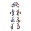 6vftC  6vfuC  6vfwC  6vg1C  6vg4C 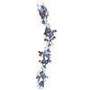 5szrS S: Starting model for refinement C: citing same article ( |
|---|---|
| Similar structure data |


-Links
 PDBj
PDBj


- Assembly
| Deposited unit | 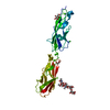
| ||||||||||||
|---|---|---|---|---|---|---|---|---|---|---|---|---|---|
| 1 |
| ||||||||||||
| Unit cell |
|
-Components
-Protein , 1 types, 1 molecules A
| #1: Protein | Mass: 24599.246 Da / Num. of mol.: 1 Source method: isolated from a genetically manipulated source Source: (gene. exp.) Homo sapiens (human) / Gene: PCDH8 / Cell line (production host): HEK293 / Production host: Homo sapiens (human) / References: UniProt: O95206 |
|---|
-Sugars , 2 types, 2 molecules 
| #2: Polysaccharide | alpha-D-mannopyranose-(1-6)-beta-D-mannopyranose-(1-4)-2-acetamido-2-deoxy-beta-D-glucopyranose-(1- ...alpha-D-mannopyranose-(1-6)-beta-D-mannopyranose-(1-4)-2-acetamido-2-deoxy-beta-D-glucopyranose-(1-4)-[alpha-L-fucopyranose-(1-6)]2-acetamido-2-deoxy-beta-D-glucopyranose Source method: isolated from a genetically manipulated source |
|---|---|
| #3: Sugar | ChemComp-MAN /  Type: D-saccharide, alpha linking / Mass: 180.156 Da / Num. of mol.: 1 Type: D-saccharide, alpha linking / Mass: 180.156 Da / Num. of mol.: 1Source method: isolated from a genetically manipulated source Formula: C6H12O6 |
-Non-polymers , 4 types, 13 molecules 


| #4: Chemical | ChemComp-CA /  Mass: 40.078 Da / Num. of mol.: 4 Mass: 40.078 Da / Num. of mol.: 4Source method: isolated from a genetically manipulated source Formula: Ca #5: Chemical | ChemComp-EDO / |  Mass: 62.068 Da / Num. of mol.: 1 Mass: 62.068 Da / Num. of mol.: 1Source method: isolated from a genetically manipulated source Formula: C2H6O2 #6: Chemical | ChemComp-CL / |  Mass: 35.453 Da / Num. of mol.: 1 / Source method: obtained synthetically / Formula: Cl Mass: 35.453 Da / Num. of mol.: 1 / Source method: obtained synthetically / Formula: Cl#7: Water | ChemComp-HOH / | Mass: 18.015 Da / Num. of mol.: 7 / Source method: isolated from a natural source / Formula: H2O |
|---|
-Details
| Has ligand of interest | N |
|---|---|
| Has protein modification | Y |
-Experimental details
-Experiment
| Experiment | Method: X-RAY DIFFRACTION / Number of used crystals: 1 |
|---|
- Sample preparation
| Crystal | Density Matthews: 2.37 Å3/Da / Density % sol: 48.11 % |
|---|---|
| Crystal grow | Temperature: 296 K / Method: vapor diffusion, hanging drop / pH: 6.5 Details: 10% PEG 20 000 (w/v), 20% (v/v) PEG 550 monomethylether, 0.03M diethyleneglycol, 0.03M triethyleneglycol, 0.03M tetraethyleneglycol, 0.03M pentaethyleneglycol, 0.1M MES/imidazole buffer pH 6.5 |
-Data collection
| Diffraction | Mean temperature: 100 K / Serial crystal experiment: N |
|---|---|
| Diffraction source | Source: SYNCHROTRON / Site: APS / Beamline: 24-ID-E / Wavelength: 0.9792 Å |
| Detector | Type: DECTRIS EIGER X 16M / Detector: PIXEL / Date: Apr 16, 2016 |
| Radiation | Protocol: SINGLE WAVELENGTH / Monochromatic (M) / Laue (L): M / Scattering type: x-ray |
| Radiation wavelength | Wavelength: 0.9792 Å / Relative weight: 1 |
| Reflection | Resolution: 2.9→40 Å / Num. obs: 5168 / % possible obs: 99.6 % / Redundancy: 3.3 % / Biso Wilson estimate: 45.4199136562 Å2 / CC1/2: 0.99 / Rmerge(I) obs: 0.13 / Rpim(I) all: 0.08 / Rrim(I) all: 0.16 / Net I/σ(I): 8.2 |
| Reflection shell | Resolution: 2.9→3.07 Å / Redundancy: 2.3 % / Rmerge(I) obs: 0.57 / Mean I/σ(I) obs: 1.7 / Num. unique obs: 803 / CC1/2: 0.71 / Rpim(I) all: 0.44 / Rrim(I) all: 0.72 / % possible all: 99.1 |
- Processing
| Software |
| |||||||||||||||||||||||||||||||||||||||||||||||||||||||||||||||||||||||||||
|---|---|---|---|---|---|---|---|---|---|---|---|---|---|---|---|---|---|---|---|---|---|---|---|---|---|---|---|---|---|---|---|---|---|---|---|---|---|---|---|---|---|---|---|---|---|---|---|---|---|---|---|---|---|---|---|---|---|---|---|---|---|---|---|---|---|---|---|---|---|---|---|---|---|---|---|---|
| Refinement | Method to determine structure: MOLECULAR REPLACEMENT Starting model: 5SZR Resolution: 2.9→19.9 Å / SU ML: 0.3763 / Cross valid method: FREE R-VALUE / σ(F): 1.34 / Phase error: 21.39
| |||||||||||||||||||||||||||||||||||||||||||||||||||||||||||||||||||||||||||
| Solvent computation | Shrinkage radii: 0.8 Å / VDW probe radii: 1 Å | |||||||||||||||||||||||||||||||||||||||||||||||||||||||||||||||||||||||||||
| Displacement parameters | Biso mean: 52.68 Å2 | |||||||||||||||||||||||||||||||||||||||||||||||||||||||||||||||||||||||||||
| Refinement step | Cycle: LAST / Resolution: 2.9→19.9 Å
| |||||||||||||||||||||||||||||||||||||||||||||||||||||||||||||||||||||||||||
| Refine LS restraints |
| |||||||||||||||||||||||||||||||||||||||||||||||||||||||||||||||||||||||||||
| LS refinement shell | Resolution: 2.9→3.65 Å
| |||||||||||||||||||||||||||||||||||||||||||||||||||||||||||||||||||||||||||
| Refinement TLS params. | Method: refined / Refine-ID: X-RAY DIFFRACTION
| |||||||||||||||||||||||||||||||||||||||||||||||||||||||||||||||||||||||||||
| Refinement TLS group |
|
