 Movie
Movie Controller


+ Open data

- Basic information
| Entry | Database: PDB / ID: 6vdk | ||||||||||||||||||||||||||||||||||||||||||
|---|---|---|---|---|---|---|---|---|---|---|---|---|---|---|---|---|---|---|---|---|---|---|---|---|---|---|---|---|---|---|---|---|---|---|---|---|---|---|---|---|---|---|---|
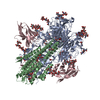
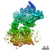
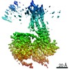
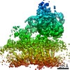
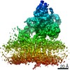
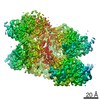
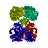
| Title | CryoEM structure of HIV-1 conserved Intasome Core | ||||||||||||||||||||||||||||||||||||||||||
 Components Components |
| ||||||||||||||||||||||||||||||||||||||||||
 Keywords Keywords | TRANSFERASE/DNA / site-specific recombination / retroviruses / integrase / integration / nucleoprotein complex / DNA complex / integrase strand transfer inhibitor / TRANSFERASE-DNA complex | ||||||||||||||||||||||||||||||||||||||||||
| Function / homology |  Function and homology information Function and homology informationHIV-1 retropepsin / symbiont-mediated activation of host apoptosis / retroviral ribonuclease H / exoribonuclease H / exoribonuclease H activity / DNA integration / viral genome integration into host DNA / establishment of integrated proviral latency / RNA-directed DNA polymerase / RNA stem-loop binding ...HIV-1 retropepsin / symbiont-mediated activation of host apoptosis / retroviral ribonuclease H / exoribonuclease H / exoribonuclease H activity / DNA integration / viral genome integration into host DNA / establishment of integrated proviral latency / RNA-directed DNA polymerase / RNA stem-loop binding / viral penetration into host nucleus / host multivesicular body / RNA-directed DNA polymerase activity / RNA-DNA hybrid ribonuclease activity / Transferases; Transferring phosphorus-containing groups; Nucleotidyltransferases / host cell / viral nucleocapsid / endonuclease activity / DNA recombination / DNA-directed DNA polymerase / aspartic-type endopeptidase activity / Hydrolases; Acting on ester bonds / host cell cytoplasm / DNA-directed DNA polymerase activity / symbiont-mediated suppression of host gene expression / viral translational frameshifting / symbiont entry into host cell / lipid binding / host cell nucleus / host cell plasma membrane / virion membrane / structural molecule activity / proteolysis / DNA binding / zinc ion binding Similarity search - Function | ||||||||||||||||||||||||||||||||||||||||||
| Biological species |   Human immunodeficiency virus 1 Human immunodeficiency virus 1 | ||||||||||||||||||||||||||||||||||||||||||
| Method | ELECTRON MICROSCOPY / single particle reconstruction / cryo EM / Resolution: 4.5 Å | ||||||||||||||||||||||||||||||||||||||||||
 Authors Authors | Li, M. / Chen, X. / Craigie, R. | ||||||||||||||||||||||||||||||||||||||||||
| Funding support |  United States, 2items United States, 2items
| ||||||||||||||||||||||||||||||||||||||||||
 Citation Citation | Journal: J Mol Biol / Year: 2020 Title: A Peptide Derived from Lens Epithelium-Derived Growth Factor Stimulates HIV-1 DNA Integration and Facilitates Intasome Structural Studies. Authors: Min Li / Xuemin Chen / Huaibin Wang / Kellie A Jurado / Alan N Engelman / Robert Craigie / Abstract: The low solubility and aggregation properties of HIV-1 integrase (IN) are major obstacles for biochemical and structural studies. The lens epithelium-derived growth factor (LEDGF) is a cellular ...The low solubility and aggregation properties of HIV-1 integrase (IN) are major obstacles for biochemical and structural studies. The lens epithelium-derived growth factor (LEDGF) is a cellular factor that binds IN and tethers preintegration complexes to chromatin before integration. The LEDGF also stimulates HIV-1 IN DNA strand transfer activity and improves its solubility in vitro. We show that these properties are conferred by a short peptide spanning residues 178 to 197 of the LEDGF that encompasses its AT-hook DNA-binding elements. The peptide stimulates HIV-1 IN activity both in trans and in cis. Fusion of the peptide to either the N- or C-terminus of IN results in maximal stimulation of concerted integration activity and greatly improves the solubility of the protein and nucleoprotein complexes of IN with viral DNA ends (intasomes). High-resolution structures of HIV-1 intasomes are required to understand the mechanism of IN strand transfer inhibitors (INSTIs), which are front-line drugs for the treatment of HIV-1, and how the virus can develop resistance to INSTIs. We have previously determined the structure of the HIV-1 strand transfer complex intasome. The improved biophysical properties of intasomes assembled with LEDGF peptide fusion IN have enabled us to determine the structure of the cleaved synaptic complex intasome, which is the direct target of INSTIs. | ||||||||||||||||||||||||||||||||||||||||||
| History |
|
- Structure visualization
| Movie |
Movie viewer |
|---|---|
| Structure viewer | Molecule: MolmilJmol/JSmol |
- Downloads & links
-Download
| PDBx/mmCIF format | 6vdk.cif.gz | 308 KB | Display | PDBx/mmCIF format |
|---|---|---|---|---|
| PDB format | pdb6vdk.ent.gz | 231.5 KB | Display | PDB format |
| PDBx/mmJSON format | 6vdk.json.gz | Tree view | PDBx/mmJSON format | |
| Others |  Other downloads Other downloads |
-Validation report
| Arichive directory | https://data.pdbj.org/pub/pdb/validation_reports/vd/6vdkftp://data.pdbj.org/pub/pdb/validation_reports/vd/6vdk | HTTPS FTP |
|---|
-Related structure data
| Related structure data | 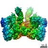 21151MC 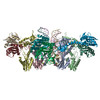 6u8qC C: citing same article ( M: map data used to model this data |
|---|---|
| Similar structure data |

-Links
 PDBj
PDBj


- Assembly
| Deposited unit | 
|
|---|---|
| 1 |
|
-Components
| #1: Protein | Mass: 39898.355 Da / Num. of mol.: 8 Source method: isolated from a genetically manipulated source Source: (gene. exp.) Human immunodeficiency virus 1 / Gene: pol / Production host: References: UniProt: F2WR39, UniProt: P12497*PLUS, Transferases; Transferring phosphorus-containing groups; Nucleotidyltransferases #2: DNA chain | Mass: 8188.271 Da / Num. of mol.: 2 / Source method: obtained synthetically / Source: (synth.) #3: DNA chain | Mass: 7773.023 Da / Num. of mol.: 2 / Source method: obtained synthetically / Source: (synth.) #4: Chemical | ChemComp-MG /   Mass: 24.305 Da / Num. of mol.: 4 / Source method: obtained synthetically / Formula: Mg / Feature type: SUBJECT OF INVESTIGATION Mass: 24.305 Da / Num. of mol.: 4 / Source method: obtained synthetically / Formula: Mg / Feature type: SUBJECT OF INVESTIGATION#5: Chemical |   Mass: 419.379 Da / Num. of mol.: 2 / Source method: obtained synthetically / Formula: C20H19F2N3O5 / Feature type: SUBJECT OF INVESTIGATION Mass: 419.379 Da / Num. of mol.: 2 / Source method: obtained synthetically / Formula: C20H19F2N3O5 / Feature type: SUBJECT OF INVESTIGATIONHas ligand of interest | Y | Has protein modification | N | |
|---|
-Experimental details
-Experiment
| Experiment | Method: ELECTRON MICROSCOPY |
|---|---|
| EM experiment | Aggregation state: PARTICLE / 3D reconstruction method: single particle reconstruction |
- Sample preparation
| Component | Name: HIV-1 conserved intasome core / Type: COMPLEX / Entity ID: #1-#3 / Source: RECOMBINANT |
|---|---|
| Source (natural) | Organism: Human immunodeficiency virus 1 |
| Source (recombinant) | Organism: |
| Buffer solution | pH: 6.2 |
| Specimen | Embedding applied: NO / Shadowing applied: NO / Staining applied: NO / Vitrification applied: YES |
| Specimen support | Details: unspecified |
| Vitrification | Cryogen name: ETHANE |
- Electron microscopy imaging
| Experimental equipment |  Model: Titan Krios / Image courtesy: FEI Company |
|---|---|
| Microscopy | Model: FEI TITAN KRIOS |
| Electron gun | Electron source:  FIELD EMISSION GUN / Accelerating voltage: 300 kV / Illumination mode: FLOOD BEAM FIELD EMISSION GUN / Accelerating voltage: 300 kV / Illumination mode: FLOOD BEAM |
| Electron lens | Mode: BRIGHT FIELD |
| Image recording | Electron dose: 75 e/Å2 / Film or detector model: GATAN K2 SUMMIT (4k x 4k) |
- Processing
| Software | Name: PHENIX / Version: 1.14_3260: / Classification: refinement | ||||||||||||||||||||||||
|---|---|---|---|---|---|---|---|---|---|---|---|---|---|---|---|---|---|---|---|---|---|---|---|---|---|
| EM software | Name: PHENIX / Category: model refinement | ||||||||||||||||||||||||
| CTF correction | Type: PHASE FLIPPING AND AMPLITUDE CORRECTION | ||||||||||||||||||||||||
| 3D reconstruction | Resolution: 4.5 Å / Resolution method: FSC 0.143 CUT-OFF / Num. of particles: 134763 / Symmetry type: POINT | ||||||||||||||||||||||||
| Refine LS restraints |
|
