 Movie
Movie Controller

[English] 日本語
 Yorodumi
- PDB-6vcw: Crystal structure of Medicago truncatula S-adenosylmethionine Syn... -
+ Open data

- Basic information
| Entry | Database: PDB / ID: 6vcw | ||||||
|---|---|---|---|---|---|---|---|
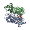
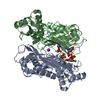
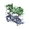
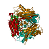
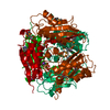
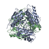
| Title | Crystal structure of Medicago truncatula S-adenosylmethionine Synthase 3A (MtMAT3A) | ||||||
 Components Components | S-adenosylmethionine synthase | ||||||
 Keywords Keywords | TRANSFERASE / methionine adenosyltransferase / SAM synthase | ||||||
| Function / homology |  Function and homology information Function and homology informationmethionine adenosyltransferase / methionine adenosyltransferase activity / S-adenosylmethionine biosynthetic process / one-carbon metabolic process / ATP binding / metal ion binding / cytosol Similarity search - Function | ||||||
| Biological species |  | ||||||
| Method |  X-RAY DIFFRACTION / SYNCHROTRON / MOLECULAR REPLACEMENT / Resolution: 1.4 Å X-RAY DIFFRACTION / SYNCHROTRON / MOLECULAR REPLACEMENT / Resolution: 1.4 Å | ||||||
 Authors Authors | Sekula, B. / Ruszkowski, M. / Dauter, Z. | ||||||
| Funding support |  United States, 1items United States, 1items
| ||||||
 Citation Citation | Journal: Int.J.Biol.Macromol. / Year: 2020 Title: S-adenosylmethionine synthases in plants: Structural characterization of type I and II isoenzymes from Arabidopsis thaliana and Medicago truncatula. Authors: Sekula, B. / Ruszkowski, M. / Dauter, Z. | ||||||
| History |
|
- Structure visualization
| Structure viewer | Molecule: MolmilJmol/JSmol |
|---|
- Downloads & links
-Download
| PDBx/mmCIF format | 6vcw.cif.gz | 346.4 KB | Display | PDBx/mmCIF format |
|---|---|---|---|---|
| PDB format | pdb6vcw.ent.gz | 279.1 KB | Display | PDB format |
| PDBx/mmJSON format | 6vcw.json.gz | Tree view | PDBx/mmJSON format | |
| Others |  Other downloads Other downloads |
-Validation report
| Arichive directory | https://data.pdbj.org/pub/pdb/validation_reports/vc/6vcwftp://data.pdbj.org/pub/pdb/validation_reports/vc/6vcw | HTTPS FTP |
|---|
-Related structure data
| Related structure data | 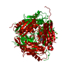 6vcxC  6vcyC 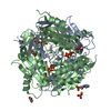 6vczC  6vd0C 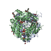 6vd1C  6vd2C 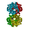 5a1iS S: Starting model for refinement C: citing same article ( |
|---|---|
| Similar structure data |


-Links
 PDBj
PDBj- Assembly
| Deposited unit | 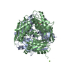
| ||||||||
|---|---|---|---|---|---|---|---|---|---|
| 1 |
| ||||||||
| Unit cell |
|
-Components
| #1: Protein | Mass: 42984.812 Da / Num. of mol.: 2 Source method: isolated from a genetically manipulated source Source: (gene. exp.)  #2: Chemical | ChemComp-MG /   Mass: 24.305 Da / Num. of mol.: 4 / Source method: obtained synthetically / Formula: Mg / Feature type: SUBJECT OF INVESTIGATION Mass: 24.305 Da / Num. of mol.: 4 / Source method: obtained synthetically / Formula: Mg / Feature type: SUBJECT OF INVESTIGATION#3: Chemical |   Mass: 35.453 Da / Num. of mol.: 2 / Source method: obtained synthetically / Formula: Cl Mass: 35.453 Da / Num. of mol.: 2 / Source method: obtained synthetically / Formula: Cl#4: Chemical | ChemComp-EDO / |   Mass: 62.068 Da / Num. of mol.: 1 / Source method: obtained synthetically / Formula: C2H6O2 Mass: 62.068 Da / Num. of mol.: 1 / Source method: obtained synthetically / Formula: C2H6O2#5: Water | ChemComp-HOH / |  Mass: 18.015 Da / Num. of mol.: 965 / Source method: isolated from a natural source / Formula: H2O Mass: 18.015 Da / Num. of mol.: 965 / Source method: isolated from a natural source / Formula: H2OHas ligand of interest | Y | |
|---|
-Experimental details
-Experiment
| Experiment | Method: X-RAY DIFFRACTION / Number of used crystals: 1 |
|---|
- Sample preparation
| Crystal | Density Matthews: 2.06 Å3/Da / Density % sol: 40.28 % |
|---|---|
| Crystal grow | Temperature: 292 K / Method: vapor diffusion, hanging drop / pH: 8 Details: 0.01 M CoCl2, 0.2 M MgCl2, 0.1 M Bis-Tris propane at pH 8.0, 6.25% PEG 3350, 6.25% PEG 4000, 6.25% PEG 2000, 6.25% mmPEG 5000 |
-Data collection
| Diffraction | Mean temperature: 100 K / Serial crystal experiment: N |
|---|---|
| Diffraction source | Source: SYNCHROTRON / Site: APS / Beamline: 19-ID / Wavelength: 0.9792 Å |
| Detector | Type: DECTRIS PILATUS 6M / Detector: PIXEL / Date: Nov 15, 2017 |
| Radiation | Monochromator: Si (111) / Protocol: SINGLE WAVELENGTH / Monochromatic (M) / Laue (L): M / Scattering type: x-ray |
| Radiation wavelength | Wavelength: 0.9792 Å / Relative weight: 1 |
| Reflection | Resolution: 1.4→48.99 Å / Num. obs: 130457 / % possible obs: 94.2 % / Observed criterion σ(I): -3 / Redundancy: 4.79 % / Rmerge(I) obs: 0.066 / Net I/σ(I): 14.48 |
| Reflection shell | Resolution: 1.4→1.48 Å / Redundancy: 4.67 % / Rmerge(I) obs: 0.762 / Mean I/σ(I) obs: 2.39 / Num. unique obs: 19394 / % possible all: 87 |
- Processing
| Software |
| |||||||||||||||||||||||||||||||||||||||||||||||||||||||||||||||||
|---|---|---|---|---|---|---|---|---|---|---|---|---|---|---|---|---|---|---|---|---|---|---|---|---|---|---|---|---|---|---|---|---|---|---|---|---|---|---|---|---|---|---|---|---|---|---|---|---|---|---|---|---|---|---|---|---|---|---|---|---|---|---|---|---|---|---|
| Refinement | Method to determine structure: MOLECULAR REPLACEMENT Starting model: 5a1i Resolution: 1.4→48.99 Å / Cor.coef. Fo:Fc: 0.986 / Cor.coef. Fo:Fc free: 0.975 / SU B: 2.49 / SU ML: 0.042 / Cross valid method: THROUGHOUT / σ(F): 0 / ESU R: 0.052 / ESU R Free: 0.053 Details: HYDROGENS HAVE BEEN ADDED IN THE RIDING POSITIONS U VALUES : REFINED INDIVIDUALLY
| |||||||||||||||||||||||||||||||||||||||||||||||||||||||||||||||||
| Solvent computation | Ion probe radii: 0.8 Å / Shrinkage radii: 0.8 Å / VDW probe radii: 1.2 Å | |||||||||||||||||||||||||||||||||||||||||||||||||||||||||||||||||
| Displacement parameters | Biso max: 101.86 Å2 / Biso mean: 18.852 Å2 / Biso min: 8.51 Å2
| |||||||||||||||||||||||||||||||||||||||||||||||||||||||||||||||||
| Refinement step | Cycle: final / Resolution: 1.4→48.99 Å
| |||||||||||||||||||||||||||||||||||||||||||||||||||||||||||||||||
| Refine LS restraints |
| |||||||||||||||||||||||||||||||||||||||||||||||||||||||||||||||||
| LS refinement shell | Resolution: 1.4→1.433 Å / Rfactor Rfree error: 0
|
