 Movie
Movie Controller

[English] 日本語
 Yorodumi
- PDB-6rjs: Inter-dimeric interface controls function and stability of S-meth... -
+ Open data

- Basic information
| Entry | Database: PDB / ID: 6rjs | ||||||
|---|---|---|---|---|---|---|---|
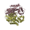
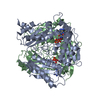
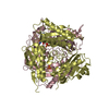
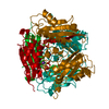
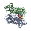
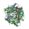
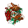
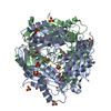
| Title | Inter-dimeric interface controls function and stability of S-methionine adenosyltransferase from U. urealiticum | ||||||
 Components Components | Methionine adenosyltransferase | ||||||
 Keywords Keywords | TRANSFERASE / synthetase | ||||||
| Function / homology | GMP Synthetase; Chain A, domain 3 - #10 / GMP Synthetase; Chain A, domain 3 / 2-Layer Sandwich / Alpha Beta / :  Function and homology information Function and homology information | ||||||
| Biological species |  Ureaplasma urealyticum serovar 7 str. ATCC 27819 (bacteria) Ureaplasma urealyticum serovar 7 str. ATCC 27819 (bacteria) | ||||||
| Method |  X-RAY DIFFRACTION / SYNCHROTRON / MOLECULAR REPLACEMENT / Resolution: 2.6 Å X-RAY DIFFRACTION / SYNCHROTRON / MOLECULAR REPLACEMENT / Resolution: 2.6 Å | ||||||
 Authors Authors | Shahar, A. / Zarivach, R. / Bershtein, S. / Kleiner, D. / Shmulevich, F. | ||||||
| Funding support |  Israel, 1items Israel, 1items
| ||||||
 Citation Citation | Journal: J.Mol.Biol. / Year: 2019 Title: The interdimeric interface controls function and stability of Ureaplasma urealiticum methionine S-adenosyltransferase. Authors: Kleiner, D. / Shmulevich, F. / Zarivach, R. / Shahar, A. / Sharon, M. / Ben-Nissan, G. / Bershtein, S. | ||||||
| History |
|
- Structure visualization
| Structure viewer | Molecule: MolmilJmol/JSmol |
|---|
- Downloads & links
-Download
| PDBx/mmCIF format | 6rjs.cif.gz | 560.8 KB | Display | PDBx/mmCIF format |
|---|---|---|---|---|
| PDB format | pdb6rjs.ent.gz | 470.6 KB | Display | PDB format |
| PDBx/mmJSON format | 6rjs.json.gz | Tree view | PDBx/mmJSON format | |
| Others |  Other downloads Other downloads |
-Validation report
| Arichive directory | https://data.pdbj.org/pub/pdb/validation_reports/rj/6rjsftp://data.pdbj.org/pub/pdb/validation_reports/rj/6rjs | HTTPS FTP |
|---|
-Related structure data
| Related structure data | 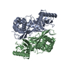 6rk5C  6rk7C  6rkaC 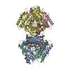 6rkcC  5t8sS S: Starting model for refinement C: citing same article ( |
|---|---|
| Similar structure data |


-Links
 PDBj
PDBj- Assembly
| Deposited unit | 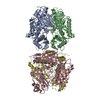
| ||||||||
|---|---|---|---|---|---|---|---|---|---|
| 1 | 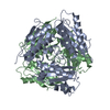
| ||||||||
| 2 | 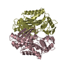
| ||||||||
| Unit cell |
|
-Components
| #1: Protein | Mass: 42189.879 Da / Num. of mol.: 4 Source method: isolated from a genetically manipulated source Source: (gene. exp.) Ureaplasma urealyticum serovar 7 str. ATCC 27819 (bacteria)Gene: metK, UUR7_0462 / Production host:  Escherichia phage EcSzw-2 (virus) / References: UniProt: B2NE58, methionine adenosyltransferase Escherichia phage EcSzw-2 (virus) / References: UniProt: B2NE58, methionine adenosyltransferase#2: Water | ChemComp-HOH / |  Mass: 18.015 Da / Num. of mol.: 83 / Source method: isolated from a natural source / Formula: H2O Mass: 18.015 Da / Num. of mol.: 83 / Source method: isolated from a natural source / Formula: H2O |
|---|
-Experimental details
-Experiment
| Experiment | Method: X-RAY DIFFRACTION / Number of used crystals: 1 |
|---|
- Sample preparation
| Crystal | Density Matthews: 2.29 Å3/Da / Density % sol: 46.36 % |
|---|---|
| Crystal grow | Temperature: 293 K / Method: vapor diffusion, sitting drop / Details: 5% Tacsimate, 0.1M Hepes pH 7.0, 10% PEG 5K |
-Data collection
| Diffraction | Mean temperature: 100 K / Serial crystal experiment: N |
|---|---|
| Diffraction source | Source: SYNCHROTRON / Site: ESRF  / Beamline: ID30B / Wavelength: 0.97625 Å / Beamline: ID30B / Wavelength: 0.97625 Å |
| Detector | Type: DECTRIS PILATUS3 S 6M / Detector: PIXEL / Date: Feb 23, 2017 |
| Radiation | Protocol: SINGLE WAVELENGTH / Monochromatic (M) / Laue (L): M / Scattering type: x-ray |
| Radiation wavelength | Wavelength: 0.97625 Å / Relative weight: 1 |
| Reflection | Resolution: 2.6→111.81 Å / Num. obs: 42461 / % possible obs: 92.02 % / Redundancy: 4.1 % / Rmerge(I) obs: 0.098 / Rpim(I) all: 0.055 / Net I/σ(I): 12.4 |
| Reflection shell | Resolution: 2.6→2.73 Å / Rmerge(I) obs: 0.648 / Num. unique obs: 12471 / CC1/2: 0.872 |
- Processing
| Software |
| ||||||||||||||||
|---|---|---|---|---|---|---|---|---|---|---|---|---|---|---|---|---|---|
| Refinement | Method to determine structure: MOLECULAR REPLACEMENT Starting model: 5T8S Resolution: 2.6→47.62 Å / Cross valid method: FREE R-VALUE
| ||||||||||||||||
| Refinement step | Cycle: LAST / Resolution: 2.6→47.62 Å
|
