 Movie
Movie Controller


+ Open data

- Basic information
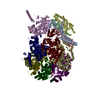
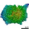
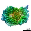
| Entry | Database: PDB / ID: 6vbv | ||||||
|---|---|---|---|---|---|---|---|
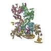
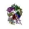
| Title | Structure of the bovine BBSome:ARL6:GTP complex | ||||||
 Components Components |
| ||||||
 Keywords Keywords | PROTEIN TRANSPORT / Cilia / ciliopathy / complex / membrane-protein transport | ||||||
| Function / homology |  Function and homology information Function and homology informationBBSome binding / establishment of anatomical structure orientation / protein transport from ciliary membrane to plasma membrane / multi-ciliated epithelial cell differentiation / receptor localization to non-motile cilium / BBSome-mediated cargo-targeting to cilium / BBSome / camera-type eye photoreceptor cell differentiation / protein localization to non-motile cilium / renal tubule development ...BBSome binding / establishment of anatomical structure orientation / protein transport from ciliary membrane to plasma membrane / multi-ciliated epithelial cell differentiation / receptor localization to non-motile cilium / BBSome-mediated cargo-targeting to cilium / BBSome / camera-type eye photoreceptor cell differentiation / protein localization to non-motile cilium / renal tubule development / smoothened binding / establishment of planar polarity / inner ear receptor cell stereocilium organization / photoreceptor connecting cilium / retina layer formation / patched binding / axonemal microtubule / membrane coat / olfactory bulb development / protein localization to cilium / establishment of epithelial cell apical/basal polarity / phosphatidylinositol-3-phosphate binding / regulation of smoothened signaling pathway / regulation of stress fiber assembly / non-motile cilium assembly / non-motile cilium / motile cilium / protein targeting to membrane / centrosome cycle / ciliary membrane / sensory perception / erythrocyte homeostasis / eating behavior / ciliary transition zone / pericentriolar material / B cell homeostasis / cilium assembly / protein polymerization / axoneme / fat cell differentiation / vesicle-mediated transport / axon guidance / protein localization to plasma membrane / intracellular protein transport / brain development / phospholipid binding / multicellular organism growth / fibrillar center / Wnt signaling pathway / centriolar satellite / sensory perception of smell / intracellular protein localization / regulation of protein localization / protein transport / gene expression / RNA polymerase II-specific DNA-binding transcription factor binding / protein-macromolecule adaptor activity / neuron projection / cilium / ciliary basal body / GTPase activity / centrosome / GTP binding / nucleoplasm / membrane / cytoplasm / cytosol Similarity search - Function | ||||||
| Biological species |  | ||||||
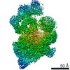
| Method | ELECTRON MICROSCOPY / single particle reconstruction / cryo EM / Resolution: 3.5 Å | ||||||
 Authors Authors | Singh, S.K. / Gui, M. / Koh, F. / Yip, M.C.J. / Brown, A. | ||||||
| Funding support |  United States, 1items United States, 1items
| ||||||
 Citation Citation | Journal: Elife / Year: 2020 Title: Structure and activation mechanism of the BBSome membrane protein trafficking complex. Authors: Sandeep K Singh / Miao Gui / Fujiet Koh / Matthew Cj Yip / Alan Brown / Abstract: Bardet-Biedl syndrome (BBS) is a currently incurable ciliopathy caused by the failure to correctly establish or maintain cilia-dependent signaling pathways. Eight proteins associated with BBS ...Bardet-Biedl syndrome (BBS) is a currently incurable ciliopathy caused by the failure to correctly establish or maintain cilia-dependent signaling pathways. Eight proteins associated with BBS assemble into the BBSome, a key regulator of the ciliary membrane proteome. We report the electron cryomicroscopy (cryo-EM) structures of the native bovine BBSome in inactive and active states at 3.1 and 3.5 Å resolution, respectively. In the active state, the BBSome is bound to an Arf-family GTPase (ARL6/BBS3) that recruits the BBSome to ciliary membranes. ARL6 recognizes a composite binding site formed by BBS1 and BBS7 that is occluded in the inactive state. Activation requires an unexpected swiveling of the β-propeller domain of BBS1, the subunit most frequently implicated in substrate recognition, which widens a central cavity of the BBSome. Structural mapping of disease-causing mutations suggests that pathogenesis results from folding defects and the disruption of autoinhibition and activation. | ||||||
| History |
|
- Structure visualization
| Movie |
Movie viewer |
|---|---|
| Structure viewer | Molecule: MolmilJmol/JSmol |
- Downloads & links
-Download
| PDBx/mmCIF format | 6vbv.cif.gz | 712.3 KB | Display | PDBx/mmCIF format |
|---|---|---|---|---|
| PDB format | pdb6vbv.ent.gz | 570.1 KB | Display | PDB format |
| PDBx/mmJSON format | 6vbv.json.gz | Tree view | PDBx/mmJSON format | |
| Others |  Other downloads Other downloads |
-Validation report
| Arichive directory | https://data.pdbj.org/pub/pdb/validation_reports/vb/6vbvftp://data.pdbj.org/pub/pdb/validation_reports/vb/6vbv | HTTPS FTP |
|---|
-Related structure data
| Related structure data | 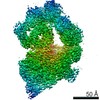 21145MC  6vbuC M: map data used to model this data C: citing same article ( |
|---|---|
| Similar structure data |


-Links
 PDBj
PDBj


- Assembly
| Deposited unit | 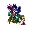
|
|---|---|
| 1 |
|
-Components
-Bardet-Biedl syndrome ... , 6 types, 6 molecules 024579
| #1: Protein | Mass: 8070.502 Da / Num. of mol.: 1 / Source method: isolated from a natural source / Source: (natural) |
|---|---|
| #3: Protein | Mass: 79911.484 Da / Num. of mol.: 1 / Source method: isolated from a natural source / Source: (natural) |
| #4: Protein | Mass: 58289.133 Da / Num. of mol.: 1 / Source method: isolated from a natural source / Source: (natural) |
| #5: Protein | Mass: 38880.984 Da / Num. of mol.: 1 / Source method: isolated from a natural source / Source: (natural) |
| #6: Protein | Mass: 80471.375 Da / Num. of mol.: 1 / Source method: isolated from a natural source / Source: (natural) |
| #8: Protein | Mass: 99230.914 Da / Num. of mol.: 1 / Source method: isolated from a natural source / Source: (natural) |
-Protein , 3 types, 3 molecules 183
| #2: Protein | Mass: 64939.141 Da / Num. of mol.: 1 / Source method: isolated from a natural source / Source: (natural) |
|---|---|
| #7: Protein | Mass: 56686.406 Da / Num. of mol.: 1 / Source method: isolated from a natural source / Source: (natural) |
| #9: Protein | Mass: 21086.486 Da / Num. of mol.: 1 Source method: isolated from a genetically manipulated source Source: (gene. exp.)  |
-Non-polymers , 2 types, 3 molecules 

| #10: Chemical |  Mass: 40.078 Da / Num. of mol.: 2 / Source method: obtained synthetically / Formula: Ca Mass: 40.078 Da / Num. of mol.: 2 / Source method: obtained synthetically / Formula: Ca#11: Chemical | ChemComp-GTP / |  Mass: 523.180 Da / Num. of mol.: 1 / Source method: obtained synthetically / Formula: C10H16N5O14P3 / Comment: GTP, energy-carrying molecule*YM Mass: 523.180 Da / Num. of mol.: 1 / Source method: obtained synthetically / Formula: C10H16N5O14P3 / Comment: GTP, energy-carrying molecule*YM |
|---|
-Details
| Has ligand of interest | N |
|---|
-Experimental details
-Experiment
| Experiment | Method: ELECTRON MICROSCOPY |
|---|---|
| EM experiment | Aggregation state: PARTICLE / 3D reconstruction method: single particle reconstruction |
- Sample preparation
| Component |
| ||||||||||||||||||||||||||||||
|---|---|---|---|---|---|---|---|---|---|---|---|---|---|---|---|---|---|---|---|---|---|---|---|---|---|---|---|---|---|---|---|
| Molecular weight | Value: 0.5 MDa / Experimental value: NO | ||||||||||||||||||||||||||||||
| Source (natural) |
| ||||||||||||||||||||||||||||||
| Source (recombinant) | Organism: | ||||||||||||||||||||||||||||||
| Buffer solution | pH: 7.5 | ||||||||||||||||||||||||||||||
| Buffer component |
| ||||||||||||||||||||||||||||||
| Specimen | Conc.: 0.7 mg/ml / Embedding applied: NO / Shadowing applied: NO / Staining applied: NO / Vitrification applied: YES | ||||||||||||||||||||||||||||||
| Specimen support | Details: unspecified | ||||||||||||||||||||||||||||||
| Vitrification | Instrument: FEI VITROBOT MARK II / Cryogen name: ETHANE / Humidity: 100 % / Chamber temperature: 293 K / Details: Grids were blotted for 2 s with a -2 offset. |
- Electron microscopy imaging
| Experimental equipment |  Model: Titan Krios / Image courtesy: FEI Company |
|---|---|
| Microscopy | Model: FEI TITAN KRIOS |
| Electron gun | Electron source:  FIELD EMISSION GUN / Accelerating voltage: 300 kV / Illumination mode: FLOOD BEAM FIELD EMISSION GUN / Accelerating voltage: 300 kV / Illumination mode: FLOOD BEAM |
| Electron lens | Mode: BRIGHT FIELD / Nominal magnification: 81000 X / Nominal defocus max: 2400 nm / Nominal defocus min: 1100 nm / Cs: 2.7 mm / C2 aperture diameter: 50 µm / Alignment procedure: COMA FREE |
| Specimen holder | Cryogen: NITROGEN / Specimen holder model: FEI TITAN KRIOS AUTOGRID HOLDER |
| Image recording | Average exposure time: 4 sec. / Electron dose: 56 e/Å2 / Film or detector model: GATAN K3 BIOQUANTUM (6k x 4k) / Num. of grids imaged: 2 / Num. of real images: 9408 |
| EM imaging optics | Energyfilter name: GIF Bioquantum / Energyfilter slit width: 25 eV |
- Processing
| EM software |
| ||||||||||||||||||||||||||||||||||||||||||||||||||
|---|---|---|---|---|---|---|---|---|---|---|---|---|---|---|---|---|---|---|---|---|---|---|---|---|---|---|---|---|---|---|---|---|---|---|---|---|---|---|---|---|---|---|---|---|---|---|---|---|---|---|---|
| CTF correction | Type: PHASE FLIPPING AND AMPLITUDE CORRECTION | ||||||||||||||||||||||||||||||||||||||||||||||||||
| Symmetry | Point symmetry: C1 (asymmetric) | ||||||||||||||||||||||||||||||||||||||||||||||||||
| 3D reconstruction | Resolution: 3.5 Å / Resolution method: FSC 0.143 CUT-OFF / Num. of particles: 75201 / Algorithm: BACK PROJECTION / Symmetry type: POINT | ||||||||||||||||||||||||||||||||||||||||||||||||||
| Atomic model building | B value: 43.6 / Protocol: OTHER / Space: REAL / Target criteria: Correlation coefficient Details: During refinement, the resolution limit was set to 3.5 Angstrom. Secondary structure, Ramachandran and rotamer restraints were applied during refinement. |
