 Movie
Movie Controller

[English] 日本語
 Yorodumi
- PDB-6v29: Complex of double mutant (T89V,K162T) of E. coli L-asparaginase I... -
+ Open data

- Basic information
| Entry | Database: PDB / ID: 6v29 | ||||||
|---|---|---|---|---|---|---|---|
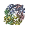
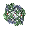
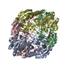
| Title | Complex of double mutant (T89V,K162T) of E. coli L-asparaginase II with L-Asp | ||||||
 Components Components | L-asparaginase 2 | ||||||
 Keywords Keywords | HYDROLASE / L-asparagine hydrolase / anti-cancer drug / inactive mutant | ||||||
| Function / homology |  Function and homology information Function and homology informationL-asparagine catabolic process / asparaginase / asparaginase activity / outer membrane-bounded periplasmic space / protein homotetramerization / periplasmic space / protein-containing complex / identical protein binding Similarity search - Function | ||||||
| Biological species |  | ||||||
| Method |  X-RAY DIFFRACTION / MOLECULAR REPLACEMENT / Resolution: 2 Å X-RAY DIFFRACTION / MOLECULAR REPLACEMENT / Resolution: 2 Å | ||||||
 Authors Authors | Lubkowski, J. / Wlodawer, A. | ||||||
 Citation Citation | Journal: Biochemistry / Year: 2020 Title: Mechanism of Catalysis by l-Asparaginase. Authors: Lubkowski, J. / Vanegas, J. / Chan, W.K. / Lorenzi, P.L. / Weinstein, J.N. / Sukharev, S. / Fushman, D. / Rempe, S. / Anishkin, A. / Wlodawer, A. | ||||||
| History |
|
- Structure visualization
| Structure viewer | Molecule: MolmilJmol/JSmol |
|---|
- Downloads & links
-Download
| PDBx/mmCIF format | 6v29.cif.gz | 265.9 KB | Display | PDBx/mmCIF format |
|---|---|---|---|---|
| PDB format | pdb6v29.ent.gz | 211.5 KB | Display | PDB format |
| PDBx/mmJSON format | 6v29.json.gz | Tree view | PDBx/mmJSON format | |
| Others |  Other downloads Other downloads |
-Validation report
| Arichive directory | https://data.pdbj.org/pub/pdb/validation_reports/v2/6v29ftp://data.pdbj.org/pub/pdb/validation_reports/v2/6v29 | HTTPS FTP |
|---|
-Related structure data
| Related structure data |  6v23C  6v24C  6v25C  6v26C  6v27C  6v28C  6v2aC  6v2bC  6v2cC  6v2gC  3ecaS S: Starting model for refinement C: citing same article ( |
|---|---|
| Similar structure data |


-Links
 PDBj
PDBj
- Assembly
| Deposited unit | 
| ||||||||
|---|---|---|---|---|---|---|---|---|---|
| 1 | 
| ||||||||
| 2 | 
| ||||||||
| Unit cell |
|
-Components
| #1: Protein | Mass: 35556.828 Da / Num. of mol.: 4 / Mutation: T89V, K162T Source method: isolated from a genetically manipulated source Source: (gene. exp.) Strain: K12 / Gene: ansB, b2957, JW2924 / Plasmid: pET-22b(+) Details (production host): ORF contains a secretion sequence, 'HHHHHH' affinity tag and sequence of doubly mutated mature EcAII Cell (production host): mesophilic bacteria / Production host: #2: Chemical | ChemComp-ASP /   Type: L-peptide linking / Mass: 133.103 Da / Num. of mol.: 4 / Source method: obtained synthetically / Formula: C4H7NO4 / Feature type: SUBJECT OF INVESTIGATION Type: L-peptide linking / Mass: 133.103 Da / Num. of mol.: 4 / Source method: obtained synthetically / Formula: C4H7NO4 / Feature type: SUBJECT OF INVESTIGATION#3: Chemical | ChemComp-GOL /   Mass: 92.094 Da / Num. of mol.: 6 / Source method: obtained synthetically / Formula: C3H8O3 Mass: 92.094 Da / Num. of mol.: 6 / Source method: obtained synthetically / Formula: C3H8O3#4: Water | ChemComp-HOH / |  Mass: 18.015 Da / Num. of mol.: 1018 / Source method: isolated from a natural source / Formula: H2O Mass: 18.015 Da / Num. of mol.: 1018 / Source method: isolated from a natural source / Formula: H2OHas ligand of interest | Y | Has protein modification | Y | |
|---|
-Experimental details
-Experiment
| Experiment | Method: X-RAY DIFFRACTION / Number of used crystals: 1 |
|---|
- Sample preparation
| Crystal | Density Matthews: 2.1 Å3/Da / Density % sol: 41.43 % |
|---|---|
| Crystal grow | Temperature: 293 K / Method: vapor diffusion, hanging drop / pH: 6.2 Details: Crystals grown at 0.17 M sodium citrate (pH 6) and 17-18% (w/v) PEG3350. Soaked for 1-2 minutes (empirically determined for each crystal) in equivalent solution with 0.025% (w/v) ...Details: Crystals grown at 0.17 M sodium citrate (pH 6) and 17-18% (w/v) PEG3350. Soaked for 1-2 minutes (empirically determined for each crystal) in equivalent solution with 0.025% (w/v) glutaraldehyde. Finally transferred and soaked for 10-20 sec in solution containing 40% (w/v) PEG3350, 5 mM L-L-Asp, and 0.17 M sodium citrate (pH 6.2) |
-Data collection
| Diffraction | Mean temperature: 100 K / Serial crystal experiment: N | |||||||||||||||||||||||||||||||||||||||||||||||||||||||||||||||||||||||||||||||||||||||||||||||||||||||||||||||||||||||||||||||||||||||||||||||||||||||||||||||||||||||||||||||||||||||||||||
|---|---|---|---|---|---|---|---|---|---|---|---|---|---|---|---|---|---|---|---|---|---|---|---|---|---|---|---|---|---|---|---|---|---|---|---|---|---|---|---|---|---|---|---|---|---|---|---|---|---|---|---|---|---|---|---|---|---|---|---|---|---|---|---|---|---|---|---|---|---|---|---|---|---|---|---|---|---|---|---|---|---|---|---|---|---|---|---|---|---|---|---|---|---|---|---|---|---|---|---|---|---|---|---|---|---|---|---|---|---|---|---|---|---|---|---|---|---|---|---|---|---|---|---|---|---|---|---|---|---|---|---|---|---|---|---|---|---|---|---|---|---|---|---|---|---|---|---|---|---|---|---|---|---|---|---|---|---|---|---|---|---|---|---|---|---|---|---|---|---|---|---|---|---|---|---|---|---|---|---|---|---|---|---|---|---|---|---|---|---|---|
| Diffraction source | Source: ROTATING ANODE / Type: RIGAKU MICROMAX-007 HF / Wavelength: 1.5418 Å | |||||||||||||||||||||||||||||||||||||||||||||||||||||||||||||||||||||||||||||||||||||||||||||||||||||||||||||||||||||||||||||||||||||||||||||||||||||||||||||||||||||||||||||||||||||||||||||
| Detector | Type: DECTRIS EIGER X 4M / Detector: PIXEL / Date: May 22, 2018 | |||||||||||||||||||||||||||||||||||||||||||||||||||||||||||||||||||||||||||||||||||||||||||||||||||||||||||||||||||||||||||||||||||||||||||||||||||||||||||||||||||||||||||||||||||||||||||||
| Radiation | Protocol: SINGLE WAVELENGTH / Monochromatic (M) / Laue (L): M / Scattering type: x-ray | |||||||||||||||||||||||||||||||||||||||||||||||||||||||||||||||||||||||||||||||||||||||||||||||||||||||||||||||||||||||||||||||||||||||||||||||||||||||||||||||||||||||||||||||||||||||||||||
| Radiation wavelength | Wavelength: 1.5418 Å / Relative weight: 1 | |||||||||||||||||||||||||||||||||||||||||||||||||||||||||||||||||||||||||||||||||||||||||||||||||||||||||||||||||||||||||||||||||||||||||||||||||||||||||||||||||||||||||||||||||||||||||||||
| Reflection | Resolution: 2→40 Å / Num. obs: 79279 / % possible obs: 99.1 % / Redundancy: 3.2 % / Rmerge(I) obs: 0.06 / Rpim(I) all: 0.039 / Rrim(I) all: 0.072 / Χ2: 0.879 / Net I/σ(I): 10.7 / Num. measured all: 256387 | |||||||||||||||||||||||||||||||||||||||||||||||||||||||||||||||||||||||||||||||||||||||||||||||||||||||||||||||||||||||||||||||||||||||||||||||||||||||||||||||||||||||||||||||||||||||||||||
| Reflection shell | Diffraction-ID: 1
|
- Processing
| Software |
| ||||||||||||||||||||||||||||||||||||||||||||||||||||||||||||
|---|---|---|---|---|---|---|---|---|---|---|---|---|---|---|---|---|---|---|---|---|---|---|---|---|---|---|---|---|---|---|---|---|---|---|---|---|---|---|---|---|---|---|---|---|---|---|---|---|---|---|---|---|---|---|---|---|---|---|---|---|---|
| Refinement | Method to determine structure: MOLECULAR REPLACEMENT Starting model: 3eca Resolution: 2→26 Å / Cor.coef. Fo:Fc: 0.972 / Cor.coef. Fo:Fc free: 0.945 / SU B: 4.024 / SU ML: 0.109 / SU R Cruickshank DPI: 0.162 / Cross valid method: THROUGHOUT / σ(F): 0 / ESU R: 0.162 / ESU R Free: 0.155 Details: HYDROGENS HAVE BEEN ADDED IN THE RIDING POSITIONS U VALUES : REFINED INDIVIDUALLY
| ||||||||||||||||||||||||||||||||||||||||||||||||||||||||||||
| Solvent computation | Ion probe radii: 0.8 Å / Shrinkage radii: 0.8 Å / VDW probe radii: 1.2 Å | ||||||||||||||||||||||||||||||||||||||||||||||||||||||||||||
| Displacement parameters | Biso max: 117.12 Å2 / Biso mean: 30.012 Å2 / Biso min: 12.73 Å2
| ||||||||||||||||||||||||||||||||||||||||||||||||||||||||||||
| Refinement step | Cycle: final / Resolution: 2→26 Å
| ||||||||||||||||||||||||||||||||||||||||||||||||||||||||||||
| Refine LS restraints |
| ||||||||||||||||||||||||||||||||||||||||||||||||||||||||||||
| LS refinement shell | Resolution: 2.001→2.052 Å / Rfactor Rfree error: 0 / Total num. of bins used: 20
|
