 Movie
Movie Controller

[English] 日本語
 Yorodumi
- PDB-6v09: Crystal structure of human recombinant Beta-2 glycoprotein I shor... -
+ Open data

- Basic information
| Entry | Database: PDB / ID: 6v09 | |||||||||
|---|---|---|---|---|---|---|---|---|---|---|
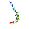
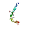
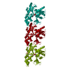
| Title | Crystal structure of human recombinant Beta-2 glycoprotein I short tag (ST-B2GPI) | |||||||||
 Components Components | Beta-2-glycoprotein 1 | |||||||||
 Keywords Keywords | BLOOD CLOTTING / PLASMA GLYCOPROTEIN / COAGULATION / INNATE IMMUNE SYSTEM / AUTOIMMUNITY / THROMBOSIS / SUSHI DOMAIN | |||||||||
| Function / homology |  Function and homology information Function and homology informationpositive regulation of triglyceride metabolic process / lipoprotein lipase activator activity / triglyceride transport / chylomicron remodeling / platelet dense granule lumen / lipase binding / blood coagulation, intrinsic pathway / very-low-density lipoprotein particle remodeling / negative regulation of myeloid cell apoptotic process / regulation of fibrinolysis ...positive regulation of triglyceride metabolic process / lipoprotein lipase activator activity / triglyceride transport / chylomicron remodeling / platelet dense granule lumen / lipase binding / blood coagulation, intrinsic pathway / very-low-density lipoprotein particle remodeling / negative regulation of myeloid cell apoptotic process / regulation of fibrinolysis / chylomicron / high-density lipoprotein particle / very-low-density lipoprotein particle / negative regulation of endothelial cell migration / plasminogen activation / negative regulation of endothelial cell proliferation / negative regulation of smooth muscle cell apoptotic process / negative regulation of blood coagulation / negative regulation of fibrinolysis / positive regulation of blood coagulation / negative regulation of angiogenesis / phospholipid binding / Platelet degranulation / heparin binding / lipid binding / cell surface / : / extracellular exosome / extracellular region / identical protein binding Similarity search - Function | |||||||||
| Biological species |  Homo sapiens (human) Homo sapiens (human) | |||||||||
| Method |  X-RAY DIFFRACTION / MOLECULAR REPLACEMENT / Resolution: 2.99 Å X-RAY DIFFRACTION / MOLECULAR REPLACEMENT / Resolution: 2.99 Å | |||||||||
 Authors Authors | Chen, Z. / Ruben, E.A. / Planer, W. / Chinnaraj, M. / Zuo, X. / Pengo, V. / Macor, P. / Tedesco, F. / Pozzi, N. | |||||||||
| Funding support |  United States, 1items United States, 1items
| |||||||||
 Citation Citation | Journal: J.Biol.Chem. / Year: 2020 Title: The J-elongated conformation of beta2-glycoprotein I predominates in solution: implications for our understanding of antiphospholipid syndrome. Authors: Ruben, E. / Planer, W. / Chinnaraj, M. / Chen, Z. / Zuo, X. / Pengo, V. / De Filippis, V. / Alluri, R.K. / McCrae, K.R. / Macor, P. / Tedesco, F. / Pozzi, N. #1: Journal: EMBO J. / Year: 1999Title: CRYSTAL STRUCTURE OF HUMAN BETA2-GLYCOPROTEIN I: IMPLICATIONS FOR PHOSPHOLIPID BINDING AND THE ANTIPHOSPHOLIPID SYNDROME. Authors: Schwarzenbacher, R. / Zeth, K. / Diederichs, K. / Gries, A. / Kostner, G.M. / Laggner, P. / Prassl, R. | |||||||||
| History |
|
- Structure visualization
| Structure viewer | Molecule: MolmilJmol/JSmol |
|---|
- Downloads & links
-Download
| PDBx/mmCIF format | 6v09.cif.gz | 87.1 KB | Display | PDBx/mmCIF format |
|---|---|---|---|---|
| PDB format | pdb6v09.ent.gz | 64.1 KB | Display | PDB format |
| PDBx/mmJSON format | 6v09.json.gz | Tree view | PDBx/mmJSON format | |
| Others |  Other downloads Other downloads |
-Validation report
| Arichive directory | https://data.pdbj.org/pub/pdb/validation_reports/v0/6v09ftp://data.pdbj.org/pub/pdb/validation_reports/v0/6v09 | HTTPS FTP |
|---|
-Related structure data
| Related structure data | 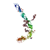 6v06C 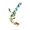 6v08C  1c1zS S: Starting model for refinement C: citing same article ( |
|---|---|
| Similar structure data |


-Links
 PDBj
PDBj

- Assembly
| Deposited unit | 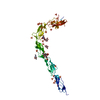
| |||||||||
|---|---|---|---|---|---|---|---|---|---|---|
| 1 |
| |||||||||
| Unit cell |
| |||||||||
| Components on special symmetry positions |
|
-Components
| #1: Protein | Mass: 37668.086 Da / Num. of mol.: 1 Source method: isolated from a genetically manipulated source Details: SLAFW is disordered in the electron densities of coordinates. EDQVDPRLIDGK is the purification tag. EDQVDPRLI is disordered in the electron densities of coordinates Source: (gene. exp.) Homo sapiens (human) / Gene: APOH, B2G1 / Cell line (production host): HEK293 / Production host: Homo sapiens (human) / References: UniProt: P02749 | ||||||||||
|---|---|---|---|---|---|---|---|---|---|---|---|
| #2: Polysaccharide | Source method: isolated from a genetically manipulated source #3: Sugar |   Type: D-saccharide, beta linking / Mass: 221.208 Da / Num. of mol.: 2 Type: D-saccharide, beta linking / Mass: 221.208 Da / Num. of mol.: 2Source method: isolated from a genetically manipulated source Formula: C8H15NO6 #4: Chemical | ChemComp-SO4 /   Mass: 96.063 Da / Num. of mol.: 26 Mass: 96.063 Da / Num. of mol.: 26Source method: isolated from a genetically manipulated source Formula: SO4 #5: Water | ChemComp-HOH / |  Mass: 18.015 Da / Num. of mol.: 15 / Source method: isolated from a natural source / Formula: H2O Mass: 18.015 Da / Num. of mol.: 15 / Source method: isolated from a natural source / Formula: H2OHas ligand of interest | N | Has protein modification | Y | |
-Experimental details
-Experiment
| Experiment | Method: X-RAY DIFFRACTION / Number of used crystals: 1 |
|---|
- Sample preparation
| Crystal | Density Matthews: 10.87 Å3/Da / Density % sol: 88.7 % |
|---|---|
| Crystal grow | Temperature: 277 K / Method: vapor diffusion, hanging drop / pH: 7.5 Details: 100 mM HEPES, 1.5 M AmSO4, 20 mM CaCl2 and 2% glycerol |
-Data collection
| Diffraction | Mean temperature: 100 K / Serial crystal experiment: N |
|---|---|
| Diffraction source | Source: ROTATING ANODE / Type: Cu FINE FOCUS / Wavelength: 1.5418 Å |
| Detector | Type: RIGAKU RAXIS IV++ / Detector: IMAGE PLATE / Date: Apr 2, 2015 |
| Radiation | Protocol: SINGLE WAVELENGTH / Monochromatic (M) / Laue (L): M / Scattering type: x-ray |
| Radiation wavelength | Wavelength: 1.5418 Å / Relative weight: 1 |
| Reflection | Resolution: 2.99→116.98 Å / Num. obs: 31543 / % possible obs: 98.8 % / Redundancy: 5 % / Rmerge(I) obs: 0.095 / Net I/σ(I): 13.7 |
| Reflection shell | Resolution: 3→3.05 Å / Redundancy: 3.7 % / Rmerge(I) obs: 0.461 / Mean I/σ(I) obs: 2.4 / Num. unique obs: 1531 / % possible all: 97.7 |
- Processing
| Software |
| |||||||||||||||||||||||||||||||||||||||||||||||||||||||||||||||||||||||||||||||||||||||||||||||||||||||||||||||||||||||||||||||||||||||||||||||||
|---|---|---|---|---|---|---|---|---|---|---|---|---|---|---|---|---|---|---|---|---|---|---|---|---|---|---|---|---|---|---|---|---|---|---|---|---|---|---|---|---|---|---|---|---|---|---|---|---|---|---|---|---|---|---|---|---|---|---|---|---|---|---|---|---|---|---|---|---|---|---|---|---|---|---|---|---|---|---|---|---|---|---|---|---|---|---|---|---|---|---|---|---|---|---|---|---|---|---|---|---|---|---|---|---|---|---|---|---|---|---|---|---|---|---|---|---|---|---|---|---|---|---|---|---|---|---|---|---|---|---|---|---|---|---|---|---|---|---|---|---|---|---|---|---|---|---|
| Refinement | Method to determine structure: MOLECULAR REPLACEMENT Starting model: 1C1Z Resolution: 2.99→116.98 Å / Cor.coef. Fo:Fc: 0.918 / Cor.coef. Fo:Fc free: 0.889 / SU B: 10.795 / SU ML: 0.183 / Cross valid method: THROUGHOUT / σ(F): 0 / ESU R: 0.264 / ESU R Free: 0.23 Details: HYDROGENS HAVE BEEN ADDED IN THE RIDING POSITIONS U VALUES : REFINED INDIVIDUALLY
| |||||||||||||||||||||||||||||||||||||||||||||||||||||||||||||||||||||||||||||||||||||||||||||||||||||||||||||||||||||||||||||||||||||||||||||||||
| Solvent computation | Ion probe radii: 0.8 Å / Shrinkage radii: 0.8 Å / VDW probe radii: 1.2 Å | |||||||||||||||||||||||||||||||||||||||||||||||||||||||||||||||||||||||||||||||||||||||||||||||||||||||||||||||||||||||||||||||||||||||||||||||||
| Displacement parameters | Biso max: 197.31 Å2 / Biso mean: 75.41 Å2 / Biso min: 30 Å2
| |||||||||||||||||||||||||||||||||||||||||||||||||||||||||||||||||||||||||||||||||||||||||||||||||||||||||||||||||||||||||||||||||||||||||||||||||
| Refinement step | Cycle: final / Resolution: 2.99→116.98 Å
| |||||||||||||||||||||||||||||||||||||||||||||||||||||||||||||||||||||||||||||||||||||||||||||||||||||||||||||||||||||||||||||||||||||||||||||||||
| Refine LS restraints |
| |||||||||||||||||||||||||||||||||||||||||||||||||||||||||||||||||||||||||||||||||||||||||||||||||||||||||||||||||||||||||||||||||||||||||||||||||
| LS refinement shell | Resolution: 2.99→3.06 Å
|
