 Movie
Movie Controller

[English] 日本語
 Yorodumi
- PDB-1c1z: CRYSTAL STRUCTURE OF HUMAN BETA-2-GLYCOPROTEIN-I (APOLIPOPROTEIN-H) -
+ Open data

- Basic information
| Entry | Database: PDB / ID: 1c1z | |||||||||
|---|---|---|---|---|---|---|---|---|---|---|
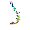
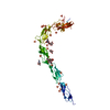
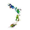
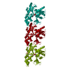
| Title | CRYSTAL STRUCTURE OF HUMAN BETA-2-GLYCOPROTEIN-I (APOLIPOPROTEIN-H) | |||||||||
 Components Components | BETA2-GLYCOPROTEIN-I | |||||||||
 Keywords Keywords | SIGNALING PROTEIN / GLYCOPROTEIN / SHORT CONSENSUS REPEAT / SCR / SUSHI DOMAIN / COMPLEMENT CONTROL PROTEIN MODULE / CCP | |||||||||
| Function / homology |  Function and homology information Function and homology informationpositive regulation of triglyceride metabolic process / lipoprotein lipase activator activity / triglyceride transport / chylomicron remodeling / platelet dense granule lumen / lipase binding / blood coagulation, intrinsic pathway / very-low-density lipoprotein particle remodeling / negative regulation of myeloid cell apoptotic process / regulation of fibrinolysis ...positive regulation of triglyceride metabolic process / lipoprotein lipase activator activity / triglyceride transport / chylomicron remodeling / platelet dense granule lumen / lipase binding / blood coagulation, intrinsic pathway / very-low-density lipoprotein particle remodeling / negative regulation of myeloid cell apoptotic process / regulation of fibrinolysis / chylomicron / high-density lipoprotein particle / very-low-density lipoprotein particle / negative regulation of endothelial cell migration / plasminogen activation / negative regulation of endothelial cell proliferation / negative regulation of smooth muscle cell apoptotic process / negative regulation of blood coagulation / negative regulation of fibrinolysis / positive regulation of blood coagulation / negative regulation of angiogenesis / phospholipid binding / Platelet degranulation / heparin binding / lipid binding / cell surface / : / extracellular exosome / extracellular region / identical protein binding Similarity search - Function | |||||||||
| Biological species |  Homo sapiens (human) Homo sapiens (human) | |||||||||
| Method |  X-RAY DIFFRACTION / SYNCHROTRON / MIR-MAD COMBINATION / Resolution: 2.87 Å X-RAY DIFFRACTION / SYNCHROTRON / MIR-MAD COMBINATION / Resolution: 2.87 Å | |||||||||
 Authors Authors | Schwarzenbacher, R. / Zeth, K. / Diederichs, K. / Gries, A. / Kostner, G.M. / Laggner, P. / Prassl, R. | |||||||||
 Citation Citation | Journal: EMBO J. / Year: 1999 Title: Crystal structure of human beta2-glycoprotein I: implications for phospholipid binding and the antiphospholipid syndrome. Authors: Schwarzenbacher, R. / Zeth, K. / Diederichs, K. / Gries, A. / Kostner, G.M. / Laggner, P. / Prassl, R. #1: Journal: Acta Crystallogr.,Sect.D / Year: 1998Title: Crystallization and preliminary X-ray crystallographic studies on apolipoprotein H (beta-2-glycoprotein-I) Authors: Saxena, A. / Gries, A. / Schwarzenbacher, R. / Kostner, G.M. / Laggner, P. / Prassl, R. | |||||||||
| History |
|
- Structure visualization
| Structure viewer | Molecule: MolmilJmol/JSmol |
|---|
- Downloads & links
-Download
| PDBx/mmCIF format | 1c1z.cif.gz | 82.9 KB | Display | PDBx/mmCIF format |
|---|---|---|---|---|
| PDB format | pdb1c1z.ent.gz | 62 KB | Display | PDB format |
| PDBx/mmJSON format | 1c1z.json.gz | Tree view | PDBx/mmJSON format | |
| Others |  Other downloads Other downloads |
-Validation report
| Arichive directory | https://data.pdbj.org/pub/pdb/validation_reports/c1/1c1zftp://data.pdbj.org/pub/pdb/validation_reports/c1/1c1z | HTTPS FTP |
|---|
-Related structure data
| Related structure data | |
|---|---|
| Similar structure data |


-Links
 PDBj
PDBj

- Assembly
| Deposited unit | 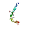
| ||||||||
|---|---|---|---|---|---|---|---|---|---|
| 1 |
| ||||||||
| Unit cell |
|
-Components
-Protein / Non-polymers , 2 types, 23 molecules A
| #1: Protein | Mass: 36313.621 Da / Num. of mol.: 1 / Source method: isolated from a natural source / Source: (natural) Homo sapiens (human) / Tissue: BLOOD / References: GenBank: 28810, UniProt: P02749*PLUS |
|---|---|
| #6: Water | ChemComp-HOH / Mass: 18.015 Da / Num. of mol.: 22 / Source method: isolated from a natural source / Formula: H2O |
-Sugars , 4 types, 4 molecules 
| #2: Polysaccharide | 2-acetamido-2-deoxy-alpha-D-glucopyranose-(1-4)-2-acetamido-2-deoxy-beta-D-glucopyranose Source method: isolated from a genetically manipulated source |
|---|---|
| #3: Polysaccharide | beta-D-mannopyranose-(1-3)-[alpha-D-mannopyranose-(1-6)]beta-D-mannopyranose-(1-4)-2-acetamido-2- ...beta-D-mannopyranose-(1-3)-[alpha-D-mannopyranose-(1-6)]beta-D-mannopyranose-(1-4)-2-acetamido-2-deoxy-alpha-D-glucopyranose-(1-4)-2-acetamido-2-deoxy-beta-D-glucopyranose Source method: isolated from a genetically manipulated source |
| #4: Polysaccharide | alpha-D-mannopyranose-(1-4)-2-acetamido-2-deoxy-beta-D-glucopyranose-(1-4)-2-acetamido-2-deoxy-beta- ...alpha-D-mannopyranose-(1-4)-2-acetamido-2-deoxy-beta-D-glucopyranose-(1-4)-2-acetamido-2-deoxy-beta-D-glucopyranose Source method: isolated from a genetically manipulated source |
| #5: Sugar | ChemComp-NAG /  Type: D-saccharide, beta linking / Mass: 221.208 Da / Num. of mol.: 1 Type: D-saccharide, beta linking / Mass: 221.208 Da / Num. of mol.: 1Source method: isolated from a genetically manipulated source Formula: C8H15NO6 |
-Details
| Has protein modification | Y |
|---|
-Experimental details
-Experiment
| Experiment | Method: X-RAY DIFFRACTION / Number of used crystals: 1 |
|---|
- Sample preparation
| Crystal | Density Matthews: 8.86 Å3/Da / Density % sol: 84 % | ||||||||||||||||||||
|---|---|---|---|---|---|---|---|---|---|---|---|---|---|---|---|---|---|---|---|---|---|
| Crystal grow | Temperature: 277 K / Method: vapor diffusion, hanging drop / pH: 7.2 Details: Ammonium sulfate, phosphate buffer, pH 7.2, VAPOR DIFFUSION, HANGING DROP, temperature 277.0K | ||||||||||||||||||||
| Crystal | *PLUS | ||||||||||||||||||||
| Crystal grow | *PLUS Temperature: 277 K / Method: vapor diffusion, sitting dropDetails: Saxena, A., (1998) Acta Crystallogr., Sect.D, 54, 1450. | ||||||||||||||||||||
| Components of the solutions | *PLUS
|
-Data collection
| Diffraction | Mean temperature: 100 K |
|---|---|
| Diffraction source | Source: SYNCHROTRON / Site: EMBL/DESY, HAMBURG  / Beamline: BW7A / Wavelength: 0.95 / Beamline: BW7A / Wavelength: 0.95 |
| Detector | Type: MARRESEARCH / Detector: IMAGE PLATE / Date: May 25, 1998 |
| Radiation | Protocol: SINGLE WAVELENGTH / Monochromatic (M) / Laue (L): M / Scattering type: x-ray |
| Radiation wavelength | Wavelength: 0.95 Å / Relative weight: 1 |
| Reflection | Resolution: 2.87→50 Å / Num. obs: 32406 / % possible obs: 93.2 % / Observed criterion σ(I): 0 / Redundancy: 3.2 % / Biso Wilson estimate: 37.9 Å2 / Rmerge(I) obs: 0.086 |
| Reflection shell | Resolution: 2.87→3.05 Å / Redundancy: 2.1 % / Rmerge(I) obs: 0.43 / % possible all: 60.1 |
| Reflection | *PLUS Highest resolution: 2.87 Å / Lowest resolution: 50 Å / Observed criterion σ(I): 0 / Redundancy: 3.2 % / Num. measured all: 103699 / Biso Wilson estimate: 37.9 Å2 |
| Reflection shell | *PLUS % possible obs: 60.1 % / Redundancy: 2.1 % / Mean I/σ(I) obs: 2.7 |
- Processing
| Software |
| ||||||||||||||||||||||||||||||||||||||||||||||||||||||||||||
|---|---|---|---|---|---|---|---|---|---|---|---|---|---|---|---|---|---|---|---|---|---|---|---|---|---|---|---|---|---|---|---|---|---|---|---|---|---|---|---|---|---|---|---|---|---|---|---|---|---|---|---|---|---|---|---|---|---|---|---|---|---|
| Refinement | Method to determine structure: MIR-MAD COMBINATION / Resolution: 2.87→50 Å / σ(F): 0 / Stereochemistry target values: ENGH & HUBER Details: SIMULATED ANNEALING REFINEMENT WITH MAXIMUM LIKELIHOOD TARGET USING AMPLITUDES, TORSION ANGLE DYNAMICS, BULK SOLVENT CORRECTION, ANISOTROPIC B- FACTOR CORRECTION AND GROUPED B-FACTOR ...Details: SIMULATED ANNEALING REFINEMENT WITH MAXIMUM LIKELIHOOD TARGET USING AMPLITUDES, TORSION ANGLE DYNAMICS, BULK SOLVENT CORRECTION, ANISOTROPIC B- FACTOR CORRECTION AND GROUPED B-FACTOR REFINEMENT SIDE CHAIN POSITIONS OF RESIDUES ARG2, ARG39, LYS59, LYS110, ARG135, GLN158, LYS177, LYS208, LYS251, LYS284, GLU285, LYS286, LYS287, LYS308, LEU313 AND PHE315 ARE POORLY DEFINED
| ||||||||||||||||||||||||||||||||||||||||||||||||||||||||||||
| Solvent computation | Solvent model: FLAT MODEL / Bsol: 33.03 Å2 / ksol: 0.342 e/Å3 | ||||||||||||||||||||||||||||||||||||||||||||||||||||||||||||
| Displacement parameters | Biso mean: 55.3 Å2
| ||||||||||||||||||||||||||||||||||||||||||||||||||||||||||||
| Refine analyze |
| ||||||||||||||||||||||||||||||||||||||||||||||||||||||||||||
| Refinement step | Cycle: LAST / Resolution: 2.87→50 Å
| ||||||||||||||||||||||||||||||||||||||||||||||||||||||||||||
| Refine LS restraints |
| ||||||||||||||||||||||||||||||||||||||||||||||||||||||||||||
| LS refinement shell | Resolution: 2.87→3.05 Å / Total num. of bins used: 6
| ||||||||||||||||||||||||||||||||||||||||||||||||||||||||||||
| Software | *PLUS Name: CNS / Classification: refinement | ||||||||||||||||||||||||||||||||||||||||||||||||||||||||||||
| Refinement | *PLUS % reflection Rfree: 5 % | ||||||||||||||||||||||||||||||||||||||||||||||||||||||||||||
| Solvent computation | *PLUS | ||||||||||||||||||||||||||||||||||||||||||||||||||||||||||||
| Displacement parameters | *PLUS | ||||||||||||||||||||||||||||||||||||||||||||||||||||||||||||
| Refine LS restraints | *PLUS
| ||||||||||||||||||||||||||||||||||||||||||||||||||||||||||||
| LS refinement shell | *PLUS Rfactor Rfree: 0.392 / % reflection Rfree: 5 % / Rfactor Rwork: 0.368 |
