 Movie
Movie Controller

[English] 日本語
 Yorodumi
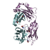
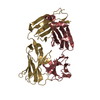
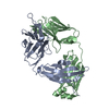
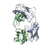
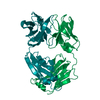
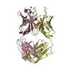
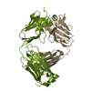
- PDB-6uum: Crystal structure of antibody 438-B11 DSS mutant (Cys98A-Cys100aA) -
+ Open data

- Basic information
| Entry | Database: PDB / ID: 6uum | ||||||
|---|---|---|---|---|---|---|---|
| Title | Crystal structure of antibody 438-B11 DSS mutant (Cys98A-Cys100aA) | ||||||
 Components Components |
| ||||||
 Keywords Keywords | IMMUNE SYSTEM / V3 glycan supersite / human antibody / anti-HIV1 neutralizing antibody / VIRAL PROTEIN | ||||||
| Function / homology | Immunoglobulins / Immunoglobulin-like / Sandwich / Mainly Beta / ACETATE ION Function and homology information Function and homology information | ||||||
| Biological species |  Homo sapiens (human) Homo sapiens (human) | ||||||
| Method |  X-RAY DIFFRACTION / SYNCHROTRON / MOLECULAR REPLACEMENT / Resolution: 2.1 Å X-RAY DIFFRACTION / SYNCHROTRON / MOLECULAR REPLACEMENT / Resolution: 2.1 Å | ||||||
 Authors Authors | Kumar, S. / Wilson, I.A. | ||||||
| Funding support |  United States, 1items United States, 1items
| ||||||
 Citation Citation | Journal: Sci Adv / Year: 2020 Title: A V H 1-69 antibody lineage from an infected Chinese donor potently neutralizes HIV-1 by targeting the V3 glycan supersite. Authors: Kumar, S. / Ju, B. / Shapero, B. / Lin, X. / Ren, L. / Zhang, L. / Li, D. / Zhou, Z. / Feng, Y. / Sou, C. / Mann, C.J. / Hao, Y. / Sarkar, A. / Hou, J. / Nunnally, C. / Hong, K. / Wang, S. / ...Authors: Kumar, S. / Ju, B. / Shapero, B. / Lin, X. / Ren, L. / Zhang, L. / Li, D. / Zhou, Z. / Feng, Y. / Sou, C. / Mann, C.J. / Hao, Y. / Sarkar, A. / Hou, J. / Nunnally, C. / Hong, K. / Wang, S. / Ge, X. / Su, B. / Landais, E. / Sok, D. / Zwick, M.B. / He, L. / Zhu, J. / Wilson, I.A. / Shao, Y. | ||||||
| History |
|
- Structure visualization
| Structure viewer | Molecule: MolmilJmol/JSmol |
|---|
- Downloads & links
-Download
| PDBx/mmCIF format | 6uum.cif.gz | 202.4 KB | Display | PDBx/mmCIF format |
|---|---|---|---|---|
| PDB format | pdb6uum.ent.gz | 156.2 KB | Display | PDB format |
| PDBx/mmJSON format | 6uum.json.gz | Tree view | PDBx/mmJSON format | |
| Others |  Other downloads Other downloads |
-Validation report
| Arichive directory | https://data.pdbj.org/pub/pdb/validation_reports/uu/6uumftp://data.pdbj.org/pub/pdb/validation_reports/uu/6uum | HTTPS FTP |
|---|
-Related structure data
| Related structure data |  6uuhC 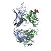 6uulC  5wl2S S: Starting model for refinement C: citing same article ( |
|---|---|
| Similar structure data |


-Links
 PDBj
PDBj

- Assembly
| Deposited unit | 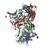
| ||||||||||||
|---|---|---|---|---|---|---|---|---|---|---|---|---|---|
| 1 | 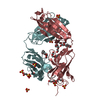
| ||||||||||||
| 2 | 
| ||||||||||||
| Unit cell |
|
-Components
-Antibody , 2 types, 4 molecules FAHB
| #1: Antibody | Mass: 23444.990 Da / Num. of mol.: 2 Source method: isolated from a genetically manipulated source Source: (gene. exp.) Homo sapiens (human) / Production host: Homo sapiens (human)#2: Antibody | Mass: 25227.479 Da / Num. of mol.: 2 Source method: isolated from a genetically manipulated source Source: (gene. exp.) Homo sapiens (human) / Production host: Homo sapiens (human) |
|---|
-Sugars , 1 types, 2 molecules 
| #4: Sugar |  Type: D-saccharide, beta linking / Mass: 221.208 Da / Num. of mol.: 2 Type: D-saccharide, beta linking / Mass: 221.208 Da / Num. of mol.: 2Source method: isolated from a genetically manipulated source Formula: C8H15NO6 |
|---|
-Non-polymers , 4 types, 559 molecules 


| #3: Chemical | ChemComp-PG4 /  Mass: 194.226 Da / Num. of mol.: 13 / Source method: obtained synthetically / Formula: C8H18O5 / Feature type: SUBJECT OF INVESTIGATION / Comment: precipitant*YM Mass: 194.226 Da / Num. of mol.: 13 / Source method: obtained synthetically / Formula: C8H18O5 / Feature type: SUBJECT OF INVESTIGATION / Comment: precipitant*YM#5: Chemical | ChemComp-SO4 /  Mass: 96.063 Da / Num. of mol.: 14 / Source method: obtained synthetically / Formula: SO4 Mass: 96.063 Da / Num. of mol.: 14 / Source method: obtained synthetically / Formula: SO4#6: Chemical |  Mass: 59.044 Da / Num. of mol.: 2 / Source method: obtained synthetically / Formula: C2H3O2 Mass: 59.044 Da / Num. of mol.: 2 / Source method: obtained synthetically / Formula: C2H3O2#7: Water | ChemComp-HOH / | Mass: 18.015 Da / Num. of mol.: 530 / Source method: isolated from a natural source / Formula: H2O |
|---|
-Details
| Has ligand of interest | Y |
|---|---|
| Has protein modification | Y |
-Experimental details
-Experiment
| Experiment | Method: X-RAY DIFFRACTION / Number of used crystals: 1 |
|---|
- Sample preparation
| Crystal | Density Matthews: 3.4 Å3/Da / Density % sol: 63.88 % |
|---|---|
| Crystal grow | Temperature: 277.15 K / Method: vapor diffusion, sitting drop Details: 0.1M sodium acetate (pH=4.16), 0.2M lithium sulfate, 40% %(v/v) PEG400 |
-Data collection
| Diffraction | Mean temperature: 100 K / Serial crystal experiment: N |
|---|---|
| Diffraction source | Source: SYNCHROTRON / Site: APS / Beamline: 23-ID-D / Wavelength: 1.033 Å |
| Detector | Type: DECTRIS PILATUS3 6M / Detector: PIXEL / Date: Oct 26, 2018 |
| Radiation | Protocol: SINGLE WAVELENGTH / Monochromatic (M) / Laue (L): M / Scattering type: x-ray |
| Radiation wavelength | Wavelength: 1.033 Å / Relative weight: 1 |
| Reflection | Resolution: 2.1→50 Å / Num. obs: 76942 / % possible obs: 99.9 % / Redundancy: 5.8 % / Biso Wilson estimate: 28.49 Å2 / CC1/2: 0.84 / Rpim(I) all: 0.07 / Rsym value: 0.17 / Net I/σ(I): 9.1 |
| Reflection shell | Resolution: 2.1→2.14 Å / Mean I/σ(I) obs: 1.2 / Num. unique obs: 3836 / CC1/2: 0.46 / Rpim(I) all: 0.52 |
- Processing
| Software |
| |||||||||||||||||||||||||||||||||||||||||||||||||||||||||||||||||||||||||||||||||||||||||||||||||||||||||
|---|---|---|---|---|---|---|---|---|---|---|---|---|---|---|---|---|---|---|---|---|---|---|---|---|---|---|---|---|---|---|---|---|---|---|---|---|---|---|---|---|---|---|---|---|---|---|---|---|---|---|---|---|---|---|---|---|---|---|---|---|---|---|---|---|---|---|---|---|---|---|---|---|---|---|---|---|---|---|---|---|---|---|---|---|---|---|---|---|---|---|---|---|---|---|---|---|---|---|---|---|---|---|---|---|---|---|
| Refinement | Method to determine structure: MOLECULAR REPLACEMENT Starting model: 5WL2 Resolution: 2.1→48.08 Å / SU ML: 0.2736 / Cross valid method: FREE R-VALUE / σ(F): 1.35 / Phase error: 26.7153 Stereochemistry target values: GeoStd + Monomer Library + CDL v1.2
| |||||||||||||||||||||||||||||||||||||||||||||||||||||||||||||||||||||||||||||||||||||||||||||||||||||||||
| Solvent computation | Shrinkage radii: 0.9 Å / VDW probe radii: 1.11 Å / Solvent model: FLAT BULK SOLVENT MODEL | |||||||||||||||||||||||||||||||||||||||||||||||||||||||||||||||||||||||||||||||||||||||||||||||||||||||||
| Displacement parameters | Biso mean: 37.2 Å2 | |||||||||||||||||||||||||||||||||||||||||||||||||||||||||||||||||||||||||||||||||||||||||||||||||||||||||
| Refinement step | Cycle: LAST / Resolution: 2.1→48.08 Å
| |||||||||||||||||||||||||||||||||||||||||||||||||||||||||||||||||||||||||||||||||||||||||||||||||||||||||
| Refine LS restraints |
| |||||||||||||||||||||||||||||||||||||||||||||||||||||||||||||||||||||||||||||||||||||||||||||||||||||||||
| LS refinement shell |
|
