 Movie
Movie Controller

[English] 日本語
 Yorodumi
- PDB-6umz: Crystal structure of photoactive yellow protein (PYP); 3-Br-p-cou... -
+ Open data

- Basic information
| Entry | Database: PDB / ID: 6umz | ||||||
|---|---|---|---|---|---|---|---|
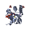
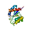
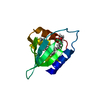
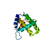
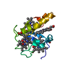
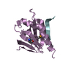
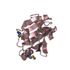
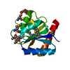
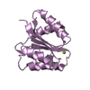
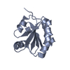
| Title | Crystal structure of photoactive yellow protein (PYP); 3-Br-p-coumaric acid chromophore | ||||||
 Components Components | Photoactive yellow protein | ||||||
 Keywords Keywords | SIGNALING PROTEIN / photoactive yellow protein | ||||||
| Function / homology |  Function and homology information Function and homology informationphotoreceptor activity / phototransduction / regulation of DNA-templated transcription / identical protein binding Similarity search - Function | ||||||
| Biological species |  Halorhodospira halophila (bacteria) Halorhodospira halophila (bacteria) | ||||||
| Method |  X-RAY DIFFRACTION / SYNCHROTRON / MOLECULAR REPLACEMENT / Resolution: 0.9 Å X-RAY DIFFRACTION / SYNCHROTRON / MOLECULAR REPLACEMENT / Resolution: 0.9 Å | ||||||
 Authors Authors | Lin, C.-Y. / Romei, M.G. / Boxer, S.G. | ||||||
| Funding support |  United States, 1items United States, 1items
| ||||||
 Citation Citation | Journal: J Photochem Photobiol A Chem / Year: 2020 Title: Structural and spectroscopic characterization of photoactive yellow protein and photoswitchable fluorescent protein constructs containing heavy atoms. Authors: Romei, M.G. / Lin, C.Y. / Boxer, S.G. | ||||||
| History |
|
- Structure visualization
| Structure viewer | Molecule: MolmilJmol/JSmol |
|---|
- Downloads & links
-Download
| PDBx/mmCIF format | 6umz.cif.gz | 96.7 KB | Display | PDBx/mmCIF format |
|---|---|---|---|---|
| PDB format | pdb6umz.ent.gz | 74.4 KB | Display | PDB format |
| PDBx/mmJSON format | 6umz.json.gz | Tree view | PDBx/mmJSON format | |
| Others |  Other downloads Other downloads |
-Validation report
| Arichive directory | https://data.pdbj.org/pub/pdb/validation_reports/um/6umzftp://data.pdbj.org/pub/pdb/validation_reports/um/6umz | HTTPS FTP |
|---|
-Related structure data
| Related structure data |  6umyC  6un0C  6un2C 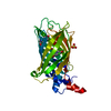 6un4C 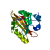 1nwzS S: Starting model for refinement C: citing same article ( |
|---|---|
| Similar structure data |
-Links
 PDBj
PDBj


- Assembly
| Deposited unit | 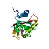
| ||||||||
|---|---|---|---|---|---|---|---|---|---|
| 1 |
| ||||||||
| Unit cell |
|
-Components
| #1: Protein | Mass: 14170.851 Da / Num. of mol.: 1 Source method: isolated from a genetically manipulated source Source: (gene. exp.) Halorhodospira halophila (bacteria) / Gene: pyp / Production host: |
|---|---|
| #2: Chemical | ChemComp-QBV / (  Mass: 243.054 Da / Num. of mol.: 1 / Source method: obtained synthetically / Formula: C9H7BrO3 / Feature type: SUBJECT OF INVESTIGATION Mass: 243.054 Da / Num. of mol.: 1 / Source method: obtained synthetically / Formula: C9H7BrO3 / Feature type: SUBJECT OF INVESTIGATION |
| #3: Water | ChemComp-HOH /  Mass: 18.015 Da / Num. of mol.: 150 / Source method: isolated from a natural source / Formula: H2O Mass: 18.015 Da / Num. of mol.: 150 / Source method: isolated from a natural source / Formula: H2O |
| Has ligand of interest | Y |
| Has protein modification | Y |
-Experimental details
-Experiment
| Experiment | Method: X-RAY DIFFRACTION / Number of used crystals: 1 |
|---|
- Sample preparation
| Crystal | Density Matthews: 1.79 Å3/Da / Density % sol: 31.19 % |
|---|---|
| Crystal grow | Temperature: 293 K / Method: vapor diffusion, hanging drop / pH: 6 Details: 20 mM potassium phosphate, pH 6.0, 1 M NaCl, 1.9 M ammonium sulfate |
-Data collection
| Diffraction | Mean temperature: 100 K / Serial crystal experiment: N |
|---|---|
| Diffraction source | Source: SYNCHROTRON / Site: SSRL / Beamline: BL9-2 / Wavelength: 0.88557 Å |
| Detector | Type: DECTRIS PILATUS 6M / Detector: PIXEL / Date: Feb 10, 2019 |
| Radiation | Protocol: SINGLE WAVELENGTH / Monochromatic (M) / Laue (L): M / Scattering type: x-ray |
| Radiation wavelength | Wavelength: 0.88557 Å / Relative weight: 1 |
| Reflection | Resolution: 0.9→33.05 Å / Num. obs: 69351 / % possible obs: 93.7 % / Redundancy: 60.1 % / Biso Wilson estimate: 8.11 Å2 / CC1/2: 1 / Rmerge(I) obs: 0.065 / Rpim(I) all: 0.008 / Rrim(I) all: 0.065 / Net I/σ(I): 39.1 / Num. measured all: 4167073 / Scaling rejects: 285 |
| Reflection shell | Resolution: 0.9→0.92 Å / Redundancy: 24.2 % / Rmerge(I) obs: 2.011 / Num. measured all: 68511 / Num. unique obs: 2834 / CC1/2: 0.656 / Rpim(I) all: 0.415 / Rrim(I) all: 2.055 / Net I/σ(I) obs: 1.9 / % possible all: 77.5 |
- Processing
| Software |
| |||||||||||||||||||||||||||||||||||||||||||||||||||||||||||||||||||||||||||||||||||||||||||||||||||||||||
|---|---|---|---|---|---|---|---|---|---|---|---|---|---|---|---|---|---|---|---|---|---|---|---|---|---|---|---|---|---|---|---|---|---|---|---|---|---|---|---|---|---|---|---|---|---|---|---|---|---|---|---|---|---|---|---|---|---|---|---|---|---|---|---|---|---|---|---|---|---|---|---|---|---|---|---|---|---|---|---|---|---|---|---|---|---|---|---|---|---|---|---|---|---|---|---|---|---|---|---|---|---|---|---|---|---|---|
| Refinement | Method to determine structure: MOLECULAR REPLACEMENT Starting model: 1NWZ Resolution: 0.9→33.05 Å / SU ML: 0.06 / Cross valid method: THROUGHOUT / σ(F): 1.34 / Phase error: 12.79
| |||||||||||||||||||||||||||||||||||||||||||||||||||||||||||||||||||||||||||||||||||||||||||||||||||||||||
| Solvent computation | Shrinkage radii: 0.9 Å / VDW probe radii: 1.11 Å | |||||||||||||||||||||||||||||||||||||||||||||||||||||||||||||||||||||||||||||||||||||||||||||||||||||||||
| Displacement parameters | Biso max: 52.38 Å2 / Biso mean: 13.02 Å2 / Biso min: 3.39 Å2 | |||||||||||||||||||||||||||||||||||||||||||||||||||||||||||||||||||||||||||||||||||||||||||||||||||||||||
| Refinement step | Cycle: final / Resolution: 0.9→33.05 Å
| |||||||||||||||||||||||||||||||||||||||||||||||||||||||||||||||||||||||||||||||||||||||||||||||||||||||||
| LS refinement shell | Refine-ID: X-RAY DIFFRACTION / Rfactor Rfree error: 0 / Total num. of bins used: 14
|
