 Movie
Movie Controller


+ Open data

- Basic information
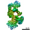
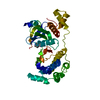
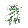
| Entry | Database: PDB / ID: 6uka | ||||||
|---|---|---|---|---|---|---|---|
| Title | Crystal structure of RHOG and ELMO complex | ||||||
 Components Components |
| ||||||
 Keywords Keywords | SIGNALING PROTEIN / RHOG / ELMO / RBD / complex / CELL ADHESION | ||||||
| Function / homology |  Function and homology information Function and homology informationPTK6 Regulates RHO GTPases, RAS GTPase and MAP kinases / regulation of ruffle assembly / RHOG GTPase cycle / Regulation of actin dynamics for phagocytic cup formation / cortical cytoskeleton organization / activation of GTPase activity / VEGFA-VEGFR2 Pathway / RHO GTPases activate KTN1 / establishment or maintenance of cell polarity / Rac protein signal transduction ...PTK6 Regulates RHO GTPases, RAS GTPase and MAP kinases / regulation of ruffle assembly / RHOG GTPase cycle / Regulation of actin dynamics for phagocytic cup formation / cortical cytoskeleton organization / activation of GTPase activity / VEGFA-VEGFR2 Pathway / RHO GTPases activate KTN1 / establishment or maintenance of cell polarity / Rac protein signal transduction / RHOG GTPase cycle / regulation of postsynapse assembly / phagocytosis / Rho protein signal transduction / GPVI-mediated activation cascade / cell projection / secretory granule membrane / actin filament organization / positive regulation of protein localization to plasma membrane / regulation of actin cytoskeleton organization / cell chemotaxis / cell motility / SH3 domain binding / receptor tyrosine kinase binding / Constitutive Signaling by Aberrant PI3K in Cancer / PIP3 activates AKT signaling / regulation of cell shape / PI5P, PP2A and IER3 Regulate PI3K/AKT Signaling / actin cytoskeleton organization / cytoplasmic vesicle / cytoskeleton / postsynapse / focal adhesion / GTPase activity / apoptotic process / positive regulation of cell population proliferation / Neutrophil degranulation / endoplasmic reticulum membrane / protein kinase binding / positive regulation of DNA-templated transcription / GTP binding / glutamatergic synapse / extracellular exosome / membrane / plasma membrane / cytosol Similarity search - Function | ||||||
| Biological species |  Homo sapiens (human) Homo sapiens (human) | ||||||
| Method |  X-RAY DIFFRACTION / MOLECULAR REPLACEMENT / Resolution: 2.4 Å X-RAY DIFFRACTION / MOLECULAR REPLACEMENT / Resolution: 2.4 Å | ||||||
 Authors Authors | Jo, C.H. / Killoran, R.C. / Smith, M.J. | ||||||
| Funding support |  Canada, 1items Canada, 1items
| ||||||
 Citation Citation | Journal: Nat Commun / Year: 2020 Title: Structure of the DOCK2-ELMO1 complex provides insights into regulation of the auto-inhibited state. Authors: Leifu Chang / Jing Yang / Chang Hwa Jo / Andreas Boland / Ziguo Zhang / Stephen H McLaughlin / Afnan Abu-Thuraia / Ryan C Killoran / Matthew J Smith / Jean-Francois Côté / David Barford /    Abstract: DOCK (dedicator of cytokinesis) proteins are multidomain guanine nucleotide exchange factors (GEFs) for RHO GTPases that regulate intracellular actin dynamics. DOCK proteins share catalytic (DOCK) ...DOCK (dedicator of cytokinesis) proteins are multidomain guanine nucleotide exchange factors (GEFs) for RHO GTPases that regulate intracellular actin dynamics. DOCK proteins share catalytic (DOCK) and membrane-associated (DOCK) domains. The structurally-related DOCK1 and DOCK2 GEFs are specific for RAC, and require ELMO (engulfment and cell motility) proteins for function. The N-terminal RAS-binding domain (RBD) of ELMO (ELMO) interacts with RHOG to modulate DOCK1/2 activity. Here, we determine the cryo-EM structures of DOCK2-ELMO1 alone, and as a ternary complex with RAC1, together with the crystal structure of a RHOG-ELMO2 complex. The binary DOCK2-ELMO1 complex adopts a closed, auto-inhibited conformation. Relief of auto-inhibition to an active, open state, due to a conformational change of the ELMO1 subunit, exposes binding sites for RAC1 on DOCK2, and RHOG and BAI GPCRs on ELMO1. Our structure explains how up-stream effectors, including DOCK2 and ELMO1 phosphorylation, destabilise the auto-inhibited state to promote an active GEF. | ||||||
| History |
|
- Structure visualization
| Structure viewer | Molecule: MolmilJmol/JSmol |
|---|
- Downloads & links
-Download
| PDBx/mmCIF format | 6uka.cif.gz | 83.1 KB | Display | PDBx/mmCIF format |
|---|---|---|---|---|
| PDB format | pdb6uka.ent.gz | 48.8 KB | Display | PDB format |
| PDBx/mmJSON format | 6uka.json.gz | Tree view | PDBx/mmJSON format | |
| Others |  Other downloads Other downloads |
-Validation report
| Arichive directory | https://data.pdbj.org/pub/pdb/validation_reports/uk/6ukaftp://data.pdbj.org/pub/pdb/validation_reports/uk/6uka | HTTPS FTP |
|---|
-Related structure data
| Related structure data | 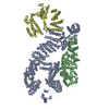 6tgbC  6tgcC  1a2bS S: Starting model for refinement C: citing same article ( |
|---|---|
| Similar structure data |


-Links
 PDBj
PDBj


- Assembly
| Deposited unit | 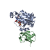
| ||||||||||||
|---|---|---|---|---|---|---|---|---|---|---|---|---|---|
| 1 |
| ||||||||||||
| Unit cell |
|
-Components
| #1: Protein | Mass: 21334.496 Da / Num. of mol.: 1 Source method: isolated from a genetically manipulated source Source: (gene. exp.) Homo sapiens (human) / Gene: RHOG, ARHG / Production host:  |
|---|---|
| #2: Protein | Mass: 9032.248 Da / Num. of mol.: 1 / Fragment: Ras-binding Domain Source method: isolated from a genetically manipulated source Source: (gene. exp.) |
| #3: Chemical | ChemComp-GNP /   Mass: 522.196 Da / Num. of mol.: 1 / Source method: isolated from a natural source / Formula: C10H17N6O13P3 / Feature type: SUBJECT OF INVESTIGATION Mass: 522.196 Da / Num. of mol.: 1 / Source method: isolated from a natural source / Formula: C10H17N6O13P3 / Feature type: SUBJECT OF INVESTIGATIONComment: GppNHp, GMPPNP, energy-carrying molecule analogue*YM |
| #4: Chemical | ChemComp-MG /   Mass: 24.305 Da / Num. of mol.: 1 / Source method: obtained synthetically / Formula: Mg Mass: 24.305 Da / Num. of mol.: 1 / Source method: obtained synthetically / Formula: Mg |
| #5: Water | ChemComp-HOH /  Mass: 18.015 Da / Num. of mol.: 126 / Source method: isolated from a natural source / Formula: H2O Mass: 18.015 Da / Num. of mol.: 126 / Source method: isolated from a natural source / Formula: H2O |
| Has ligand of interest | Y |
-Experimental details
-Experiment
| Experiment | Method: X-RAY DIFFRACTION / Number of used crystals: 1 |
|---|
- Sample preparation
| Crystal | Density Matthews: 2.12 Å3/Da / Density % sol: 42.08 % |
|---|---|
| Crystal grow | Temperature: 293 K / Method: vapor diffusion, hanging drop / Details: 0.1M CHES, 0.95M sodium citrate / PH range: 8.5 - 9.0 |
-Data collection
| Diffraction | Mean temperature: 100 K / Serial crystal experiment: N |
|---|---|
| Diffraction source | Source: SEALED TUBE / Type: BRUKER D8 QUEST / Wavelength: 1.34165 Å |
| Detector | Type: Bruker PHOTON II / Detector: PIXEL / Date: Nov 9, 2018 |
| Radiation | Protocol: SINGLE WAVELENGTH / Monochromatic (M) / Laue (L): M / Scattering type: x-ray |
| Radiation wavelength | Wavelength: 1.34165 Å / Relative weight: 1 |
| Reflection | Resolution: 2.4→27.01 Å / Num. obs: 38996 / % possible obs: 93.9 % / Redundancy: 5.7 % / Biso Wilson estimate: 17.88 Å2 / Rmerge(I) obs: 0.1131 / Net I/σ(I): 13.42 |
| Reflection shell | Resolution: 2.4→2.44 Å / Redundancy: 4.8 % / Rmerge(I) obs: 0.2591 / Mean I/σ(I) obs: 5.4 / Num. unique obs: 466 / % possible all: 89.2 |
- Processing
| Software |
| ||||||||||||||||||||||||||||||||||||||||||||||||||||||||||||||||||||||||||||||||||||||||||||||||||
|---|---|---|---|---|---|---|---|---|---|---|---|---|---|---|---|---|---|---|---|---|---|---|---|---|---|---|---|---|---|---|---|---|---|---|---|---|---|---|---|---|---|---|---|---|---|---|---|---|---|---|---|---|---|---|---|---|---|---|---|---|---|---|---|---|---|---|---|---|---|---|---|---|---|---|---|---|---|---|---|---|---|---|---|---|---|---|---|---|---|---|---|---|---|---|---|---|---|---|---|
| Refinement | Method to determine structure: MOLECULAR REPLACEMENT Starting model: 1A2B Resolution: 2.4→27.01 Å / SU ML: 0.2833 / Cross valid method: FREE R-VALUE / σ(F): 1.35 / Phase error: 24.4928
| ||||||||||||||||||||||||||||||||||||||||||||||||||||||||||||||||||||||||||||||||||||||||||||||||||
| Solvent computation | Shrinkage radii: 0.9 Å / VDW probe radii: 1.11 Å | ||||||||||||||||||||||||||||||||||||||||||||||||||||||||||||||||||||||||||||||||||||||||||||||||||
| Displacement parameters | Biso mean: 21.54 Å2 | ||||||||||||||||||||||||||||||||||||||||||||||||||||||||||||||||||||||||||||||||||||||||||||||||||
| Refinement step | Cycle: LAST / Resolution: 2.4→27.01 Å
| ||||||||||||||||||||||||||||||||||||||||||||||||||||||||||||||||||||||||||||||||||||||||||||||||||
| Refine LS restraints |
| ||||||||||||||||||||||||||||||||||||||||||||||||||||||||||||||||||||||||||||||||||||||||||||||||||
| LS refinement shell |
|
