 Movie
Movie Controller


+ Open data

- Basic information
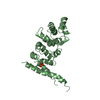
| Entry | Database: PDB / ID: 2xeq | ||||||
|---|---|---|---|---|---|---|---|
| Title | Human PatL1 C-terminal domain | ||||||
 Components Components | PAT1 HOMOLOG 1, | ||||||
 Keywords Keywords | RNA BINDING PROTEIN / MRNA DECAPPING / P-BODIES | ||||||
| Function / homology |  Function and homology information Function and homology informationpoly(G) binding / mRNA decay by 5' to 3' exoribonuclease / deadenylation-dependent decapping of nuclear-transcribed mRNA / P-body assembly / poly(U) RNA binding / P-body / PML body / cytoplasmic ribonucleoprotein granule / nuclear speck / RNA binding / cytosol Similarity search - Function | ||||||
| Biological species |  HOMO SAPIENS (human) HOMO SAPIENS (human) | ||||||
| Method |  X-RAY DIFFRACTION / SYNCHROTRON / MOLECULAR REPLACEMENT / Resolution: 3.1 Å X-RAY DIFFRACTION / SYNCHROTRON / MOLECULAR REPLACEMENT / Resolution: 3.1 Å | ||||||
 Authors Authors | Tritschler, F. / Weichenrieder, O. | ||||||
 Citation Citation | Journal: Embo J. / Year: 2010 Title: The C-Terminal Alpha-Alpha Superhelix of Pat is Required for Mrna Decapping in Metazoa. Authors: Braun, J.E. / Tritschler, F. / Haas, G. / Igreja, C. / Truffault, V. / Weichenrieder, O. / Izaurralde, E. | ||||||
| History |
| ||||||
| Remark 650 | HELIX DETERMINATION METHOD: AUTHOR PROVIDED. |
- Structure visualization
| Structure viewer | Molecule: MolmilJmol/JSmol |
|---|
- Downloads & links
-Download
| PDBx/mmCIF format | 2xeq.cif.gz | 198.2 KB | Display | PDBx/mmCIF format |
|---|---|---|---|---|
| PDB format | pdb2xeq.ent.gz | 161.1 KB | Display | PDB format |
| PDBx/mmJSON format | 2xeq.json.gz | Tree view | PDBx/mmJSON format | |
| Others |  Other downloads Other downloads |
-Validation report
| Arichive directory | https://data.pdbj.org/pub/pdb/validation_reports/xe/2xeqftp://data.pdbj.org/pub/pdb/validation_reports/xe/2xeq | HTTPS FTP |
|---|
-Related structure data
| Related structure data | 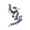 2xerC 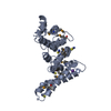 2xesSC C: citing same article ( S: Starting model for refinement |
|---|---|
| Similar structure data |


-Links
 PDBj
PDBj- Assembly
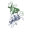
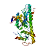
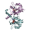
| Deposited unit | 
| ||||||||
|---|---|---|---|---|---|---|---|---|---|
| 1 | 
| ||||||||
| 2 | 
| ||||||||
| 3 | 
| ||||||||
| 4 | 
| ||||||||
| Unit cell |
|
-Components
| #1: Protein | Mass: 29604.238 Da / Num. of mol.: 4 / Fragment: C-TERMINAL DOMAIN, RESIDUES 517-767 Source method: isolated from a genetically manipulated source Source: (gene. exp.) HOMO SAPIENS (human) / Cell line: HELA / Plasmid: MODIFIED PRSFDUET-1 / Production host:  #2: Water | ChemComp-HOH / |  Mass: 18.015 Da / Num. of mol.: 8 / Source method: isolated from a natural source / Formula: H2O Mass: 18.015 Da / Num. of mol.: 8 / Source method: isolated from a natural source / Formula: H2OHas protein modification | Y | Sequence details | THE FIRST 5 AMINO ACIDS GPQDP RESULT FROM THE 5' CLONING SITE. | |
|---|
-Experimental details
-Experiment
| Experiment | Method: X-RAY DIFFRACTION / Number of used crystals: 1 |
|---|
- Sample preparation
| Crystal | Density Matthews: 2.58 Å3/Da / Density % sol: 52.4 % / Description: NONE |
|---|---|
| Crystal grow | pH: 7.8 Details: 0.1 M BIS-TRIS-PROPANE, PH 7.8, 10% PEG3350, 0.2 M NA3-CITRATE |
-Data collection
| Diffraction | Mean temperature: 100 K |
|---|---|
| Diffraction source | Source: SYNCHROTRON / Site: SLS  / Beamline: X10SA / Wavelength: 0.9794 / Beamline: X10SA / Wavelength: 0.9794 |
| Detector | Type: DECTRIS PILATUS 6M / Detector: PIXEL / Date: Dec 8, 2008 / Details: MIRRORS |
| Radiation | Monochromator: SI(111) / Protocol: SINGLE WAVELENGTH / Monochromatic (M) / Laue (L): M / Scattering type: x-ray |
| Radiation wavelength | Wavelength: 0.9794 Å / Relative weight: 1 |
| Reflection | Resolution: 3.1→43.9 Å / Num. obs: 22432 / % possible obs: 99.4 % / Observed criterion σ(I): 0 / Redundancy: 4.7 % / Biso Wilson estimate: 70.18 Å2 / Rsym value: 0.13 / Net I/σ(I): 9.1 |
| Reflection shell | Resolution: 3.1→3.18 Å / Redundancy: 4.8 % / Mean I/σ(I) obs: 1.9 / Rsym value: 0.88 / % possible all: 99.9 |
- Processing
| Software |
| |||||||||||||||||||||||||||||||||||||||||||||||||||||||||||||||||||||||||||||
|---|---|---|---|---|---|---|---|---|---|---|---|---|---|---|---|---|---|---|---|---|---|---|---|---|---|---|---|---|---|---|---|---|---|---|---|---|---|---|---|---|---|---|---|---|---|---|---|---|---|---|---|---|---|---|---|---|---|---|---|---|---|---|---|---|---|---|---|---|---|---|---|---|---|---|---|---|---|---|
| Refinement | Method to determine structure: MOLECULAR REPLACEMENT Starting model: PDB ENTRY 2XES Resolution: 3.1→43.919 Å / SU ML: 0.47 / σ(F): 1.21 / Phase error: 28.04 / Stereochemistry target values: ML Details: RESIDUES 512-514 AND 573-576 AND 704-711 AND 766-767 OF CHAIN A ARE DISORDERED. RESIDUES 512-514 AND 573-577 AND 703-710 AND 766-767 OF CHAIN B ARE DISORDERED. RESIDUES 512-514 AND 574-577 ...Details: RESIDUES 512-514 AND 573-576 AND 704-711 AND 766-767 OF CHAIN A ARE DISORDERED. RESIDUES 512-514 AND 573-577 AND 703-710 AND 766-767 OF CHAIN B ARE DISORDERED. RESIDUES 512-514 AND 574-577 AND 704-711 AND 766-767 OF CHAIN C ARE DISORDERED. RESIDUES 512-515 AND 663-670 AND 704-710 AND 766-767 OF CHAIN D ARE DISORDERED.
| |||||||||||||||||||||||||||||||||||||||||||||||||||||||||||||||||||||||||||||
| Solvent computation | Shrinkage radii: 0.9 Å / VDW probe radii: 1.11 Å / Solvent model: FLAT BULK SOLVENT MODEL / Bsol: 44.788 Å2 / ksol: 0.305 e/Å3 | |||||||||||||||||||||||||||||||||||||||||||||||||||||||||||||||||||||||||||||
| Displacement parameters | Biso mean: 78 Å2
| |||||||||||||||||||||||||||||||||||||||||||||||||||||||||||||||||||||||||||||
| Refinement step | Cycle: LAST / Resolution: 3.1→43.919 Å
| |||||||||||||||||||||||||||||||||||||||||||||||||||||||||||||||||||||||||||||
| Refine LS restraints |
| |||||||||||||||||||||||||||||||||||||||||||||||||||||||||||||||||||||||||||||
| LS refinement shell |
|
