 Movie
Movie Controller

[English] 日本語
 Yorodumi
- PDB-6uk5: Structure of SAM bound CalS10, an amino pentose methyltransferase... -
+ Open data

- Basic information
| Entry | Database: PDB / ID: 6uk5 | ||||||||||||
|---|---|---|---|---|---|---|---|---|---|---|---|---|---|
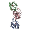
| Title | Structure of SAM bound CalS10, an amino pentose methyltransferase from Micromonospora echinaspora involved in calicheamicin biosynthesis | ||||||||||||
 Components Components | CalS10 | ||||||||||||
 Keywords Keywords | TRANSFERASE / methyltransferase / natural product / calicheamicin biosynthesis | ||||||||||||
| Function / homology | : / Methyltransferase domain 25 / Methyltransferase domain / methyltransferase activity / S-adenosyl-L-methionine-dependent methyltransferase superfamily / ACETATE ION / DI(HYDROXYETHYL)ETHER / S-ADENOSYLMETHIONINE / CalS10 Function and homology information Function and homology information | ||||||||||||
| Biological species |  Micromonospora echinospora (bacteria) Micromonospora echinospora (bacteria) | ||||||||||||
| Method |  X-RAY DIFFRACTION / SYNCHROTRON / MOLECULAR REPLACEMENT / molecular replacement / Resolution: 2.6 Å X-RAY DIFFRACTION / SYNCHROTRON / MOLECULAR REPLACEMENT / molecular replacement / Resolution: 2.6 Å | ||||||||||||
 Authors Authors | Alvarado, S.K. / Miller, M.D. / Xu, W. / Wang, Z. / Van Lanen, S.G. / Thorson, J.S. / Phillips Jr., G.N. | ||||||||||||
| Funding support |  United States, 3items United States, 3items
| ||||||||||||
 Citation Citation | Journal: To Be Published Title: Structure of SAM bound CalS10, an amino pentose methyltransferase from Micromonospora echinaspora involved in calicheamicin biosynthesis Authors: Alvarado, S.K. / Miller, M.D. / Xu, W. / Wang, Z. / Van Lanen, S.G. / Thorson, J.S. / Phillips Jr., G.N. | ||||||||||||
| History |
|
- Structure visualization
| Structure viewer | Molecule: MolmilJmol/JSmol |
|---|
- Downloads & links
-Download
| PDBx/mmCIF format | 6uk5.cif.gz | 230.9 KB | Display | PDBx/mmCIF format |
|---|---|---|---|---|
| PDB format | pdb6uk5.ent.gz | 150 KB | Display | PDB format |
| PDBx/mmJSON format | 6uk5.json.gz | Tree view | PDBx/mmJSON format | |
| Others |  Other downloads Other downloads |
-Validation report
| Arichive directory | https://data.pdbj.org/pub/pdb/validation_reports/uk/6uk5ftp://data.pdbj.org/pub/pdb/validation_reports/uk/6uk5 | HTTPS FTP |
|---|
-Related structure data
| Related structure data |  3bxoS S: Starting model for refinement |
|---|---|
| Similar structure data |


-Links
 PDBj
PDBj
- Assembly
| Deposited unit | 
| ||||||||||||||||||||||||||||||||||||||||||||||||||||||||||||
|---|---|---|---|---|---|---|---|---|---|---|---|---|---|---|---|---|---|---|---|---|---|---|---|---|---|---|---|---|---|---|---|---|---|---|---|---|---|---|---|---|---|---|---|---|---|---|---|---|---|---|---|---|---|---|---|---|---|---|---|---|---|
| 1 |
| ||||||||||||||||||||||||||||||||||||||||||||||||||||||||||||
| Unit cell |
| ||||||||||||||||||||||||||||||||||||||||||||||||||||||||||||
| Noncrystallographic symmetry (NCS) | NCS domain:
NCS domain segments:
|
-Components
-Protein , 1 types, 2 molecules AB
| #1: Protein | Mass: 28631.318 Da / Num. of mol.: 2 Source method: isolated from a genetically manipulated source Source: (gene. exp.) Micromonospora echinospora (bacteria) / Gene: calS10 / Production host: |
|---|
-Non-polymers , 5 types, 34 molecules 


| #2: Chemical |  Mass: 106.120 Da / Num. of mol.: 2 / Source method: obtained synthetically / Formula: C4H10O3 Mass: 106.120 Da / Num. of mol.: 2 / Source method: obtained synthetically / Formula: C4H10O3#3: Chemical |  Mass: 398.437 Da / Num. of mol.: 2 / Source method: obtained synthetically / Formula: C15H22N6O5S / Feature type: SUBJECT OF INVESTIGATION Mass: 398.437 Da / Num. of mol.: 2 / Source method: obtained synthetically / Formula: C15H22N6O5S / Feature type: SUBJECT OF INVESTIGATION#4: Chemical |  Mass: 59.044 Da / Num. of mol.: 2 / Source method: obtained synthetically / Formula: C2H3O2 Mass: 59.044 Da / Num. of mol.: 2 / Source method: obtained synthetically / Formula: C2H3O2#5: Chemical |  Mass: 238.278 Da / Num. of mol.: 2 / Source method: obtained synthetically / Formula: C10H22O6 / Comment: precipitant*YM Mass: 238.278 Da / Num. of mol.: 2 / Source method: obtained synthetically / Formula: C10H22O6 / Comment: precipitant*YM#6: Water | ChemComp-HOH / | Mass: 18.015 Da / Num. of mol.: 26 / Source method: isolated from a natural source / Formula: H2O |
|---|
-Details
| Has ligand of interest | Y |
|---|
-Experimental details
-Experiment
| Experiment | Method: X-RAY DIFFRACTION / Number of used crystals: 1 |
|---|
- Sample preparation
| Crystal | Density Matthews: 3.66 Å3/Da / Density % sol: 66.41 % |
|---|---|
| Crystal grow | Temperature: 298 K / Method: vapor diffusion, sitting drop / pH: 5.5 Details: 0.1M sodium citrate pH5.5, 24% PEG 400, 0.28M ammonium acetate |
-Data collection
| Diffraction | Mean temperature: 100 K / Serial crystal experiment: N | ||||||||||||||||||||||||||||||
|---|---|---|---|---|---|---|---|---|---|---|---|---|---|---|---|---|---|---|---|---|---|---|---|---|---|---|---|---|---|---|---|
| Diffraction source | Source: SYNCHROTRON / Site: APS / Beamline: 21-ID-D / Wavelength: 1.078 Å | ||||||||||||||||||||||||||||||
| Detector | Type: DECTRIS EIGER X 9M / Detector: PIXEL / Date: Apr 15, 2018 / Details: Bimorph K-B pair | ||||||||||||||||||||||||||||||
| Radiation | Monochromator: Si(111) / Protocol: SINGLE WAVELENGTH / Monochromatic (M) / Laue (L): M / Scattering type: x-ray | ||||||||||||||||||||||||||||||
| Radiation wavelength | Wavelength: 1.078 Å / Relative weight: 1 | ||||||||||||||||||||||||||||||
| Reflection | Resolution: 2.6→66.71 Å / Num. obs: 25648 / % possible obs: 100 % / Redundancy: 17.9 % / CC1/2: 0.999 / Rmerge(I) obs: 0.148 / Rpim(I) all: 0.036 / Rrim(I) all: 0.153 / Net I/σ(I): 12.2 / Num. measured all: 459637 / Scaling rejects: 2080 | ||||||||||||||||||||||||||||||
| Reflection shell | Diffraction-ID: 1
|
-Phasing
| Phasing | Method: molecular replacement | |||||||||
|---|---|---|---|---|---|---|---|---|---|---|
| Phasing MR |
|
- Processing
| Software |
| |||||||||||||||||||||||||||||||||||||||||||||||||||||||||||||||||||||||||||||
|---|---|---|---|---|---|---|---|---|---|---|---|---|---|---|---|---|---|---|---|---|---|---|---|---|---|---|---|---|---|---|---|---|---|---|---|---|---|---|---|---|---|---|---|---|---|---|---|---|---|---|---|---|---|---|---|---|---|---|---|---|---|---|---|---|---|---|---|---|---|---|---|---|---|---|---|---|---|---|
| Refinement | Method to determine structure: MOLECULAR REPLACEMENT Starting model: 3bxo Resolution: 2.6→47.15 Å / SU ML: 0.3649 / Cross valid method: FREE R-VALUE / σ(F): 1.52 / Phase error: 28.3666 Stereochemistry target values: GeoStd + Monomer Library + CDL v1.2
| |||||||||||||||||||||||||||||||||||||||||||||||||||||||||||||||||||||||||||||
| Solvent computation | Shrinkage radii: 0.9 Å / VDW probe radii: 1.11 Å / Solvent model: FLAT BULK SOLVENT MODEL | |||||||||||||||||||||||||||||||||||||||||||||||||||||||||||||||||||||||||||||
| Displacement parameters | Biso mean: 62.13 Å2 | |||||||||||||||||||||||||||||||||||||||||||||||||||||||||||||||||||||||||||||
| Refinement step | Cycle: LAST / Resolution: 2.6→47.15 Å
| |||||||||||||||||||||||||||||||||||||||||||||||||||||||||||||||||||||||||||||
| Refine LS restraints |
| |||||||||||||||||||||||||||||||||||||||||||||||||||||||||||||||||||||||||||||
| LS refinement shell |
|
