 Movie
Movie Controller

[English] 日本語
 Yorodumi
- PDB-6uig: Crystal structure of human monoclonal antibody H7.200 in complex ... -
+ Open data

- Basic information
| Entry | Database: PDB / ID: 6uig | ||||||
|---|---|---|---|---|---|---|---|
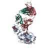
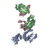
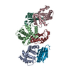
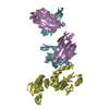
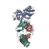
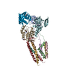
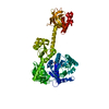
| Title | Crystal structure of human monoclonal antibody H7.200 in complex with H7N9 hemagglutinin HA1 | ||||||
 Components Components |
| ||||||
 Keywords Keywords | ANTIVIRAL PROTEIN / influenza A / monoclonal antibody | ||||||
| Function / homology |  Function and homology information Function and homology informationviral budding from plasma membrane / clathrin-dependent endocytosis of virus by host cell / host cell surface receptor binding / fusion of virus membrane with host plasma membrane / fusion of virus membrane with host endosome membrane / viral envelope / virion attachment to host cell / host cell plasma membrane / virion membrane / membrane / metal ion binding Similarity search - Function | ||||||
| Biological species |   Influenza A virus Influenza A virus Homo sapiens (human) Homo sapiens (human) | ||||||
| Method |  X-RAY DIFFRACTION / SYNCHROTRON / MOLECULAR REPLACEMENT / Resolution: 3.2 Å X-RAY DIFFRACTION / SYNCHROTRON / MOLECULAR REPLACEMENT / Resolution: 3.2 Å | ||||||
 Authors Authors | Dong, J. / Crowe, J.E. | ||||||
| Funding support |  United States, 1items United States, 1items
| ||||||
 Citation Citation | Journal: J.Clin.Invest. / Year: 2020 Title: Anti-influenza H7 human antibody targets antigenic site in hemagglutinin head domain interface. Authors: Dong, J. / Gilchuk, I. / Li, S. / Irving, R. / Goff, M.T. / Turner, H.L. / Ward, A.B. / Carnahan, R.H. / Crowe Jr., J.E. | ||||||
| History |
|
- Structure visualization
| Structure viewer | Molecule: MolmilJmol/JSmol |
|---|
- Downloads & links
-Download
| PDBx/mmCIF format | 6uig.cif.gz | 522.2 KB | Display | PDBx/mmCIF format |
|---|---|---|---|---|
| PDB format | pdb6uig.ent.gz | 429.8 KB | Display | PDB format |
| PDBx/mmJSON format | 6uig.json.gz | Tree view | PDBx/mmJSON format | |
| Others |  Other downloads Other downloads |
-Validation report
| Arichive directory | https://data.pdbj.org/pub/pdb/validation_reports/ui/6uigftp://data.pdbj.org/pub/pdb/validation_reports/ui/6uig | HTTPS FTP |
|---|
-Related structure data
| Related structure data | 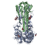 4n5jS S: Starting model for refinement |
|---|---|
| Similar structure data |


-Links
 PDBj
PDBj


- Assembly
| Deposited unit | 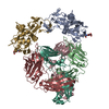
| ||||||||
|---|---|---|---|---|---|---|---|---|---|
| 1 | 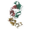
| ||||||||
| 2 | 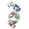
| ||||||||
| Unit cell |
|
-Components
| #1: Protein | Mass: 33755.918 Da / Num. of mol.: 2 Source method: isolated from a genetically manipulated source Source: (gene. exp.) Influenza A virus / Strain: A/Shanghai/2013(H7N9) / Gene: HA / Production host: Homo sapiens (human) / References: UniProt: A0A4Y5QYN9, UniProt: R4NN21*PLUS#2: Antibody | Mass: 24268.238 Da / Num. of mol.: 2 Source method: isolated from a genetically manipulated source Source: (gene. exp.) Homo sapiens (human) / Production host: Homo sapiens (human)#3: Antibody | Mass: 23362.854 Da / Num. of mol.: 2 Source method: isolated from a genetically manipulated source Source: (gene. exp.) Homo sapiens (human) / Production host: Homo sapiens (human)#4: Sugar |   Type: D-saccharide, beta linking / Mass: 221.208 Da / Num. of mol.: 2 Type: D-saccharide, beta linking / Mass: 221.208 Da / Num. of mol.: 2Source method: isolated from a genetically manipulated source Formula: C8H15NO6 Has ligand of interest | N | Has protein modification | Y | |
|---|
-Experimental details
-Experiment
| Experiment | Method: X-RAY DIFFRACTION / Number of used crystals: 1 |
|---|
- Sample preparation
| Crystal | Density Matthews: 4.31 Å3/Da / Density % sol: 71.45 % |
|---|---|
| Crystal grow | Temperature: 291.15 K / Method: vapor diffusion, sitting drop Details: 0.1 M sodium citrate pH 6.0, 10% isopropanol, 22% PEG 400 |
-Data collection
| Diffraction | Mean temperature: 100 K / Serial crystal experiment: N |
|---|---|
| Diffraction source | Source: SYNCHROTRON / Site: APS / Beamline: 21-ID-G / Wavelength: 0.97857 Å |
| Detector | Type: RAYONIX MX-300 / Detector: CCD / Date: Oct 11, 2018 |
| Radiation | Protocol: SINGLE WAVELENGTH / Monochromatic (M) / Laue (L): M / Scattering type: x-ray |
| Radiation wavelength | Wavelength: 0.97857 Å / Relative weight: 1 |
| Reflection | Resolution: 3.2→49.821 Å / Num. obs: 47601 / % possible obs: 99.9 % / Redundancy: 5.5 % / Rmerge(I) obs: 0.097 / Net I/σ(I): 11.3 |
| Reflection shell | Resolution: 3.2→3.37 Å / Rmerge(I) obs: 0.607 / Num. unique obs: 6843 |
- Processing
| Software |
| ||||||||||||||||||||||||||||||||||||||||||||||||||||||||||||||||||||||||||||||||||||||||||||||||||||||||||||
|---|---|---|---|---|---|---|---|---|---|---|---|---|---|---|---|---|---|---|---|---|---|---|---|---|---|---|---|---|---|---|---|---|---|---|---|---|---|---|---|---|---|---|---|---|---|---|---|---|---|---|---|---|---|---|---|---|---|---|---|---|---|---|---|---|---|---|---|---|---|---|---|---|---|---|---|---|---|---|---|---|---|---|---|---|---|---|---|---|---|---|---|---|---|---|---|---|---|---|---|---|---|---|---|---|---|---|---|---|---|
| Refinement | Method to determine structure: MOLECULAR REPLACEMENT Starting model: 4N5J Resolution: 3.2→49.821 Å / SU ML: 0.4 / Cross valid method: THROUGHOUT / σ(F): 1.34 / Phase error: 24.67 / Stereochemistry target values: ML
| ||||||||||||||||||||||||||||||||||||||||||||||||||||||||||||||||||||||||||||||||||||||||||||||||||||||||||||
| Solvent computation | Shrinkage radii: 0.9 Å / VDW probe radii: 1.11 Å / Solvent model: FLAT BULK SOLVENT MODEL | ||||||||||||||||||||||||||||||||||||||||||||||||||||||||||||||||||||||||||||||||||||||||||||||||||||||||||||
| Displacement parameters | Biso max: 174.14 Å2 / Biso mean: 88.0847 Å2 / Biso min: 30 Å2 | ||||||||||||||||||||||||||||||||||||||||||||||||||||||||||||||||||||||||||||||||||||||||||||||||||||||||||||
| Refinement step | Cycle: final / Resolution: 3.2→49.821 Å
| ||||||||||||||||||||||||||||||||||||||||||||||||||||||||||||||||||||||||||||||||||||||||||||||||||||||||||||
| LS refinement shell | Refine-ID: X-RAY DIFFRACTION / Rfactor Rfree error: 0
| ||||||||||||||||||||||||||||||||||||||||||||||||||||||||||||||||||||||||||||||||||||||||||||||||||||||||||||
| Refinement TLS params. | Method: refined / Origin x: 39.3364 Å / Origin y: -38.6148 Å / Origin z: 29.3136 Å
| ||||||||||||||||||||||||||||||||||||||||||||||||||||||||||||||||||||||||||||||||||||||||||||||||||||||||||||
| Refinement TLS group |
|
